Scaffolds with viable tissue
a technology of tissue and scaffolding, applied in the field of tissue implants having viable tissue, can solve the problems of inconvenient use, high labor intensity, and inconvenient maintenance, and achieve the effect of stimulating cell growth
- Summary
- Abstract
- Description
- Claims
- Application Information
AI Technical Summary
Benefits of technology
Problems solved by technology
Method used
Image

Examples
example 1
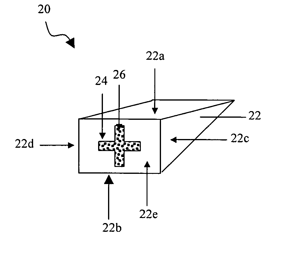
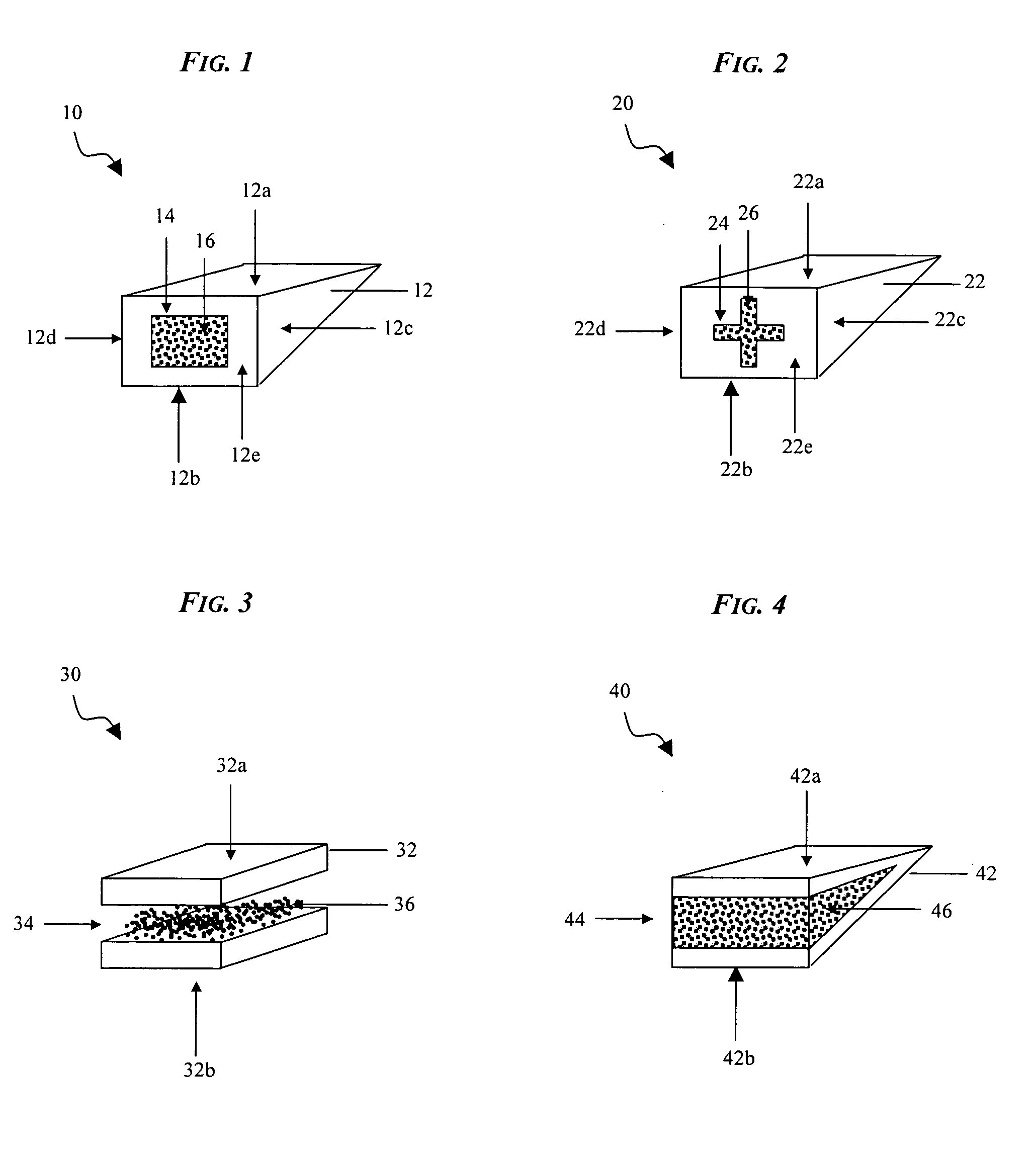
[0083] Cellular migration and new matrix formation of bovine minced meniscal tissue (BMT) into a small intestine submucosa (SIS) tissue scaffold was evaluated and compared. Meniscal tissue was harvested from the white and red-white zone of an adult bovine menisci, and the tissue was minced to form a viable tissue source. A scaffold was prepared from a small intestine submucosa (SIS), and a slit was made in the scaffold to form a pocket. The minced tissue was loaded at 20 mg / cm2 into the SIS scaffold through the slit to form a composite implant. The composite implant was placed in a pocket created in the hemithorax through one skin incision of a mouse. Tacking sutures of 5-0 Ethibond Excel® were used to tack the skin to musculature around each composite implant to prevent subcutaneous migration.
[0084] After 4 weeks, the composite implant was prepared for histological evaluation by fixing the implant in 10% buffered formalin, sectioning the implant to form samples, and staining the s...
example 2
[0086] Cellular migration and new matrix formation of bovine minced meniscal tissue (BMT) into a small intestine submucosa (SIS) tissue scaffold was evaluated and compared. Meniscal tissue was harvested from the white and red-white zone of adult bovine menisci, and the tissue was minced to form a viable tissue source. A scaffold was prepared from a small intestine submucosa (SIS), and a slit was made in the scaffold to form a pocket. The minced tissue was combined with platelet rich plasma (PRP), and the composition was loaded into the SIS scaffold at 20 mg / cm2 through the slit to form a composite implant. The composite implant was placed in a pocket created in the hemithorax through one skin incision of a mouse. Tacking sutures of 5-0 Ethibond Excel® were used to tack the skin to musculature around each composite implant to prevent subcutaneous migration.
[0087] After 4 weeks, the composite implant was prepared for histological evaluation by fixing the implant in 10% buffered forma...
example 3
[0088] Cellular migration and new matrix formation of bovine minced meniscal tissue (BMT) into a small intestine submucosa (SIS) tissue scaffold was evaluated and compared. Meniscal tissue was harvested from the white and red-white zone of adult bovine menisci, and the tissue was minced. PRP was added to the minced BMT. A viable tissue source was then prepared by combining the BMT and PRP with a bioresorbable polymer scaffold (referred to as “FPV”) in a 50:50 ratio. The resorbable scaffold used was a lyophilized foam scaffold (65% polyglycolic acid / 35% Polycaprolactone) reinforced with nonwoven fibers (mixture of PDS[Polydioxanone] and Vicryl). A scaffold was prepared from a small intestine submucosa (SIS), and a slit was made in the scaffold to form a pocket. The viable tissue source of FPV, BMT, and PRP was loaded at 20 mg / cm2 into the SIS scaffold through the slit to form a composite implant. The composite implant was placed in a pocket created in the hemithorax through one skin ...
PUM
 Abstract
Description
Claims
Application Information
 - R&D
- Intellectual Property
- Life Sciences
- Materials
- Tech Scout
- Unparalleled Data Quality
- Higher Quality Content
- 60% Fewer Hallucinations
Browse by: Latest US Patents, China's latest patents, Technical Efficacy Thesaurus, Application Domain, Technology Topic, Popular Technical Reports.
© 2025 PatSnap. All rights reserved.Legal|Privacy policy|Modern Slavery Act Transparency Statement|Sitemap|About US| Contact US: help@patsnap.com