
Methods and apparatus for processing image data to aid in detecting disease

a technology of image data and detection aid, which is applied in the field of methods and apparatus for processing medical image data to aid in the detection and diagnosis of disease, can solve the problems of no known treatment that can reverse the progress of the disease, and a large volume of data presented by a single ct scan, and the process of radiologists is time-consuming and labor-intensiv

Active Publication Date: 2005-05-19
GE MEDICAL SYST GLOBAL TECH CO LLC
View PDF14 Cites 55 Cited by
  • Summary
  • Abstract
  • Description
  • Claims
  • Application Information

AI Technical Summary

Benefits of technology

[0020] It will be seen that various configurations of the present invention allow the diagnosis, stating and / or prediction of COPD from medical images such as computed tomography scans. The automated features provided by the invention via personal computers and software can run with or without the supervision of an experienced physician or scientist, and allow effective disease diagnosis and treatment that can improve a patient's quality of life. Other configurations economically provide tracking of changeable parameters of either a person or an object utilizing imaging devices at a centralized location and personal computers, such as portable computers, of individuals.

Problems solved by technology

At present, there is no known treatment that can reverse the progress of the disease.
At best, the progress of the disease can only be halted.
The considerable volume of data presented by a single CT scan presents a time-consuming process for radiologists.
Fatigue is therefore a significant factor affecting sensitivity and specificity of the human reading.
In other diseases, such as emphysema, it is difficult for a radiologist to classify the extent of disease progression by only looking at the CT images.
However, one of the more prominent disease indicators is degradation of the alveoli and other tissue changes of the lung which are currently difficult to measure from CT image data.
Chronic bronchitis causes anatomical airway narrowing, which reduces lung function.
Airway modification typically begins with irritation from smoking and / or air pollution and can be caused / exacerbated by biological infection.
However, known techniques are not based on an underlying model of the disease, and the reliability of the results obtained with these techniques can be affected by variable scan parameters and scanner calibration as well as other disease pathologies.
In addition, known techniques do not provide estimates of the rate or location of tissue destruction.

Method used


Image

  • Methods and apparatus for processing image data to aid in detecting disease
  • Methods and apparatus for processing image data to aid in detecting disease
  • Methods and apparatus for processing image data to aid in detecting disease

Examples


Embodiment Construction

[0025] Example configurations of systems and processes that facilitate detecting, quantifying, staging, reporting and / or tracking of diseases are described below in detail. It will be appreciated that, with appropriate modification, other changeable parameters relating to persons or objects may also be tracked utilizing configurations of the present invention. A technical effect of the systems and processes described herein include at least one of facilitating automatic tracking of changeable parameters relating to a disease of a patient or other measurable parameters, and / or automated extraction of information relating to these parameters from a plurality of individuals or other tracked objects.

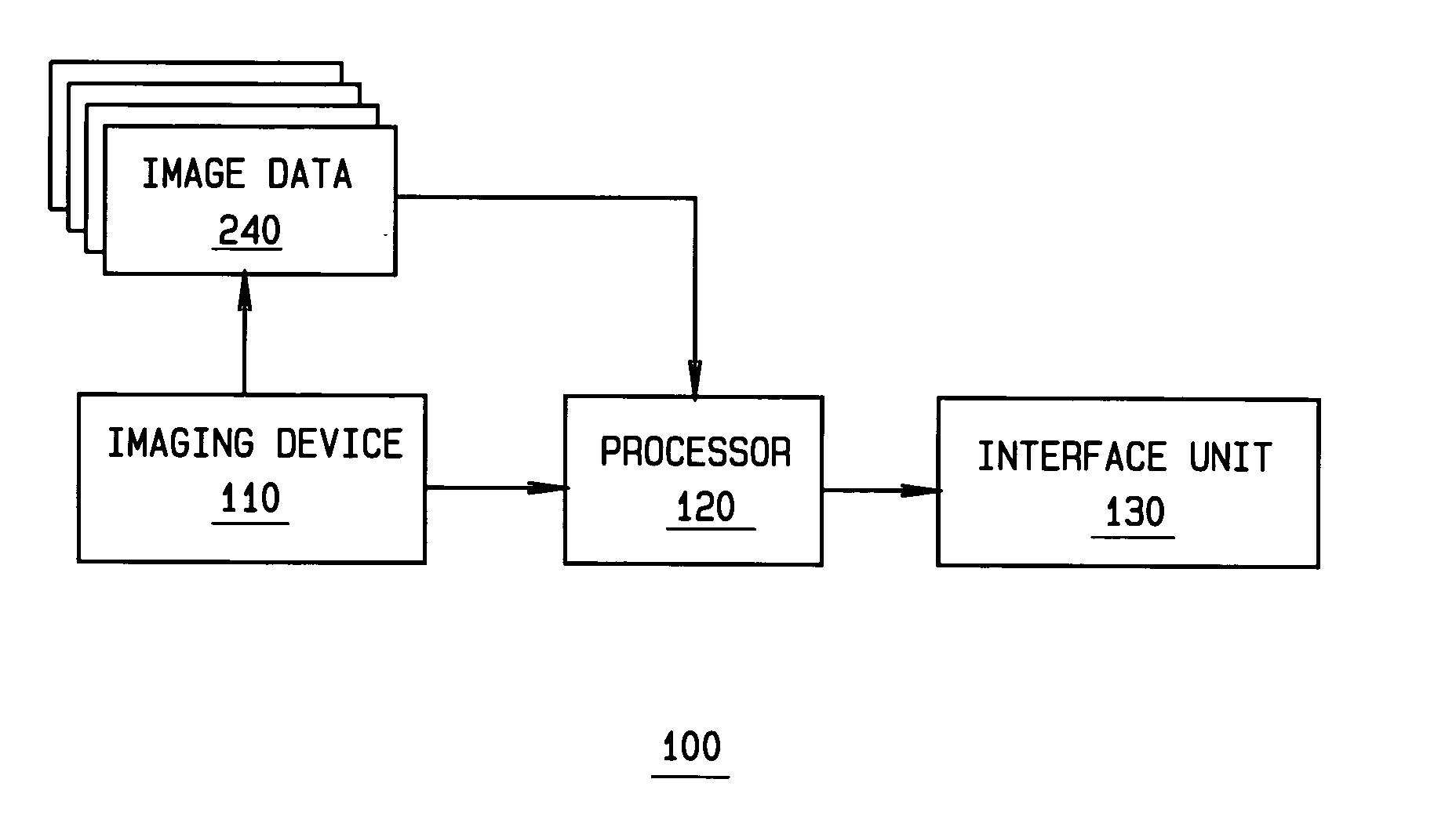
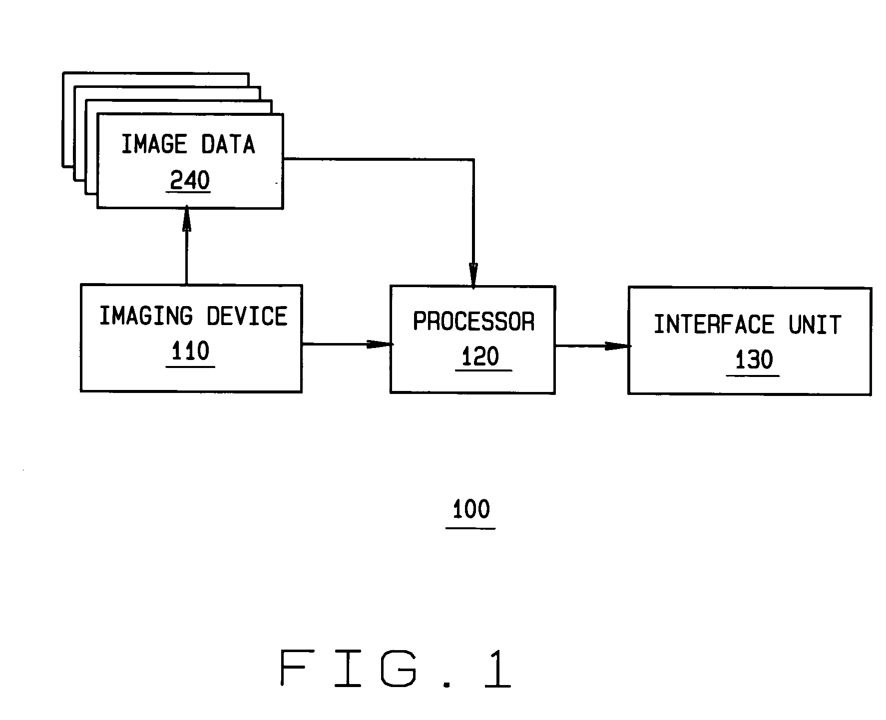
[0026] Referring to FIG. 1, a general block diagram of a system 100 for disease detection is shown. A technical effect of the system described in FIG. 1 is achieved by a user collecting and transmitting images of a patient or other object. System 100 includes an imaging device 110, which ca...


PUM


Abstract

A method for detecting, quantifying, staging, reporting, and / or tracking of a disease includes providing analysis software configured to detect, quantify, stage, report, and / or track a disease utilizing images of a patient. The analysis software is executable on a personal computer of a patient. Patients are then imaged utilizing a medical imaging apparatus and medical images of the patient produced by the imaging apparatus are downloaded to the personal computer of the patient. The imaging and downloading are repeated a plurality of times at intervals selected to provide the analysis software with sufficient images to detect, quantify, stage, report, and / or track the disease in the patient.

Description

BACKGROUND OF THE INVENTION [0001] This invention relates to methods and apparatus for processing medical image data to aid in the detection and diagnosis of disease, and more particularly, to methods and apparatus for detecting, quantifying, staging, reporting, and / or tracking of diseases such as chronic obstructive pulmonary disease. [0002] Chronic Obstructive Pulmonary Disease (COPD) is a leading cause of death in the United States and other countries. COPD has two main disease processes, namely, tissue destruction (emphysema) and airway inflammation (chronic bronchitis). At present, there is no known treatment that can reverse the progress of the disease. At best, the progress of the disease can only be halted. Thus, there is a premium placed on early disease diagnosis and treatment. With early diagnosis and effective treatment, a patient's quality of life can be improved. [0003] An x-ray chest radiograph system is the more commonly used diagnostic tool useful for the purpose of...

Claims


Application Information

Patent Timeline
IPC IPC(8): A61B5/08A61B10/00A61B6/03A61B19/00G06F17/00G06K9/00G06T7/00
CPCG06F19/321G06T2207/30061G06T7/0012G06F19/345G16H50/20G16H30/20
Inventor TUREK, MATTHEW WILLIAMMUNDY, JOSEPH LEAGRANDPAN, TONY CHI-SHAOTU, PETER HENRYMILLER, JAMES VRADENBURGKAUCIC, ROBERT AUGUST JR.WU, XIAOYESANTOS MENDONCA, PAULO RICARDO DOS
Owner GE MEDICAL SYST GLOBAL TECH CO LLC
Learn More