Methods of screening for apoptosis-controlling agents for bone anabolic therapies and uses thereof

a technology of apoptosis control and bone anabolic therapy, which is applied in the field of bone physiology, can solve the problems of glucocorticoid therapy sometimes developing collapse of the femoral head, affecting the rate of bone remodeling, and underlying uncertainty, and achieves the effect of increasing apoptosis

Inactive Publication Date: 2005-03-31
MANOLAGAS STAVROS C +3
View PDF13 Cites 3 Cited by
  • Summary
  • Abstract
  • Description
  • Claims
  • Application Information

AI Technical Summary

Benefits of technology

[0016] Intermittent PTH administration increases bone mass, but the mechanism of this effect has remained heretofore unknown. Daily PTH injections in mice either with normal bone mass or osteopenia due to defective osteoblastogenesis increased bone formation without affecting the generation of new osteoblasts. Instead, PTH did increase the life span of mature osteoblasts by preventing their apoptosis, an effect reproduced in vitro. Increasing the work performed by a cell population to augment tissue mass by suppressing apoptosis represents a novel biologic paradigm for regenerating tissues; and could provide a pharmacotherapeutic strategy for rebuilding bone in patients with established osteopenia.
[0017] Evidence is presented herein that human parathyroid hormone 1-34 [hPTH(1-34)] exerts anti-apoptotic effects on osteoblasts when administered in an intermittent fashion to mice in vivo. The present invention also provides evidence that bovine PTH(1-34) [bPTH(1-34)] prevents glucocorticoid-induced apoptosis of osteoblastic and osteocytic cells in vitro.
[0018] One object of the present invention is to provide methods for screening compounds that prevent osteoblast apoptosis, thereby stimulating bone formation and / or restoring bone in osteopenic individuals, or preventing bone loss caused by agents such as glucocorticoids.
[0019] In an embodiment of the present invention, there is provided a method of reducing the number of osteoblasts undergoing apoptosis in an individual in need of such treatment, comprising the step of: administering a therapeutic dose of human parathyroid hormone [hPTH(1-34)] to said individual, wherein administration of human parathyroid hormone [hPTH(1-34)] results in a reduction in the number of osteoblasts undergoing apoptosis, thereby reducing bone loss and / or stimulating bone formation in said individual.
[0021] In yet another embodiment of the present invention, there is provided a method of screening for compounds that decrease bone loss, comprising the steps of: (a) treating osteoblast cells with a glucocorticoid; (b) contacting said osteoblast cells with a test compound; (c) determining the number of said osteoblast cells undergoing apoptosis; and (d) comparing the number of apoptotic cells with osteoblast cells that have been treated with said glucocorticoid but were not contacted with said test compound, wherein fewer apoptotic cells following contact with said test compound than in the absence of said contact with said test compound indicates that said compound inhibits apoptosis of osteoblast cells thereby reducing bone loss.

Problems solved by technology

In addition, systemic hormones modulate the production and / or action of locally produced cytokines and growth factors, thereby influencing the rate of bone remodeling.
In addition, patients receiving long-term glucocorticoid therapy sometimes develop collapse of the femoral head (osteonecrosis), but the mechanism underlying this is uncertain.
Besides the relationship between aberrant osteoblast production and osteoporosis, it has been recently shown that a significant proportion of osteoblasts undergo apoptosis, which raises the possibility that the premature or more frequent occurrence of osteoblast apoptosis could contribute to incomplete repair of resorption cavities and loss of bone.
The mechanism of these anabolic effects, however, has not been established.
The prior art is deficient in methods of inhibiting apoptosis of osteoblasts and osteocytes.

Method used


Image

  • Methods of screening for apoptosis-controlling agents for bone anabolic therapies and uses thereof
  • Methods of screening for apoptosis-controlling agents for bone anabolic therapies and uses thereof
  • Methods of screening for apoptosis-controlling agents for bone anabolic therapies and uses thereof

Examples


example 1

In Vitro Effects of PTH

[0046] The number of osteoblasts, a critical determinant of bone formation and bone mass, depends both on the birth rate of these cells, which reflects the frequency of cell division of mesenchymal precursors, and on their life span, which reflects the timing of death by apoptosis. In vivo evidence indicates that intermittent administration of parathyroid hormone(1-34) increases bone formation and BMD in mice and that these changes are associated with decreased osteoblast and osteocyte apoptosis, but not with increased production of progenitors in the bone marrow.

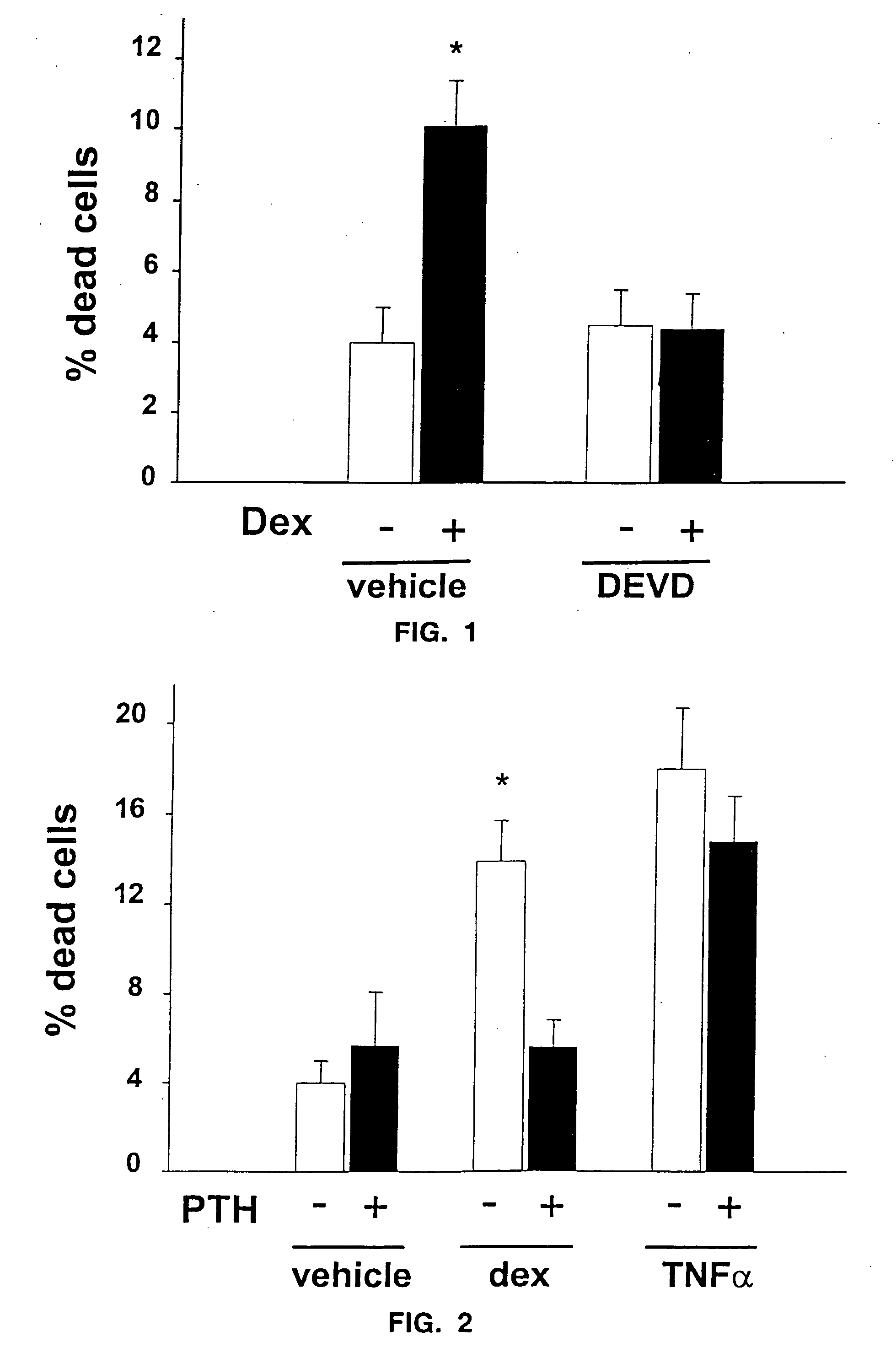
[0047] To determine the mechanism of such actions, the effects of parathyroid hormone(1-34) on the apoptosis of cultured osteoblastic cells isolated from neonatal murine calvaria and the MLO-Y4 osteocyte cell line (provided by L. Bonewald) were examined. Chromatin condensation, nuclear fragmentation, and DNA degradation—cardinal features of apoptotic cells—were monitored by microscopic examination o...

example 2

Calvaria and MLO-Y4 Cells

[0049] Osteoblastic calvaria cells (9) were cultured in αMEM (Gibco-BRL, Grand Island, N.Y.) supplemented with 10% FBS (Sigma Chemical Co., St. Louis, Mo.). Murine osteocyte-like MLO-Y4 cells stably transfected with EGFP were cultured on collagen coated plates in (MEM supplemented with 5% FBS and 5% bovine calf serum. Cultures were maintained for 6 hours in the presence of 10−7 M dexamethasone without or with preincubation for 1 hour with 10−8 M bPTH(1-34) and fixed in neutral buffered formalin. The pyknotic fragmented nuclei (arrows) typical of apoptotic cells were visualized with Hoescht 33258 fluorescent dye (Polysciences, Inc., Bayshore, N.Y.), used at a concentration of 1 μg / ml in 0.5 M NaCl, 10 mM Tris-HCl, 1 mM EDTA, pH 7.4) in osteoblastic calvaria cells, and by EGFP fluorescence in MLO-Y4 osteocytes.

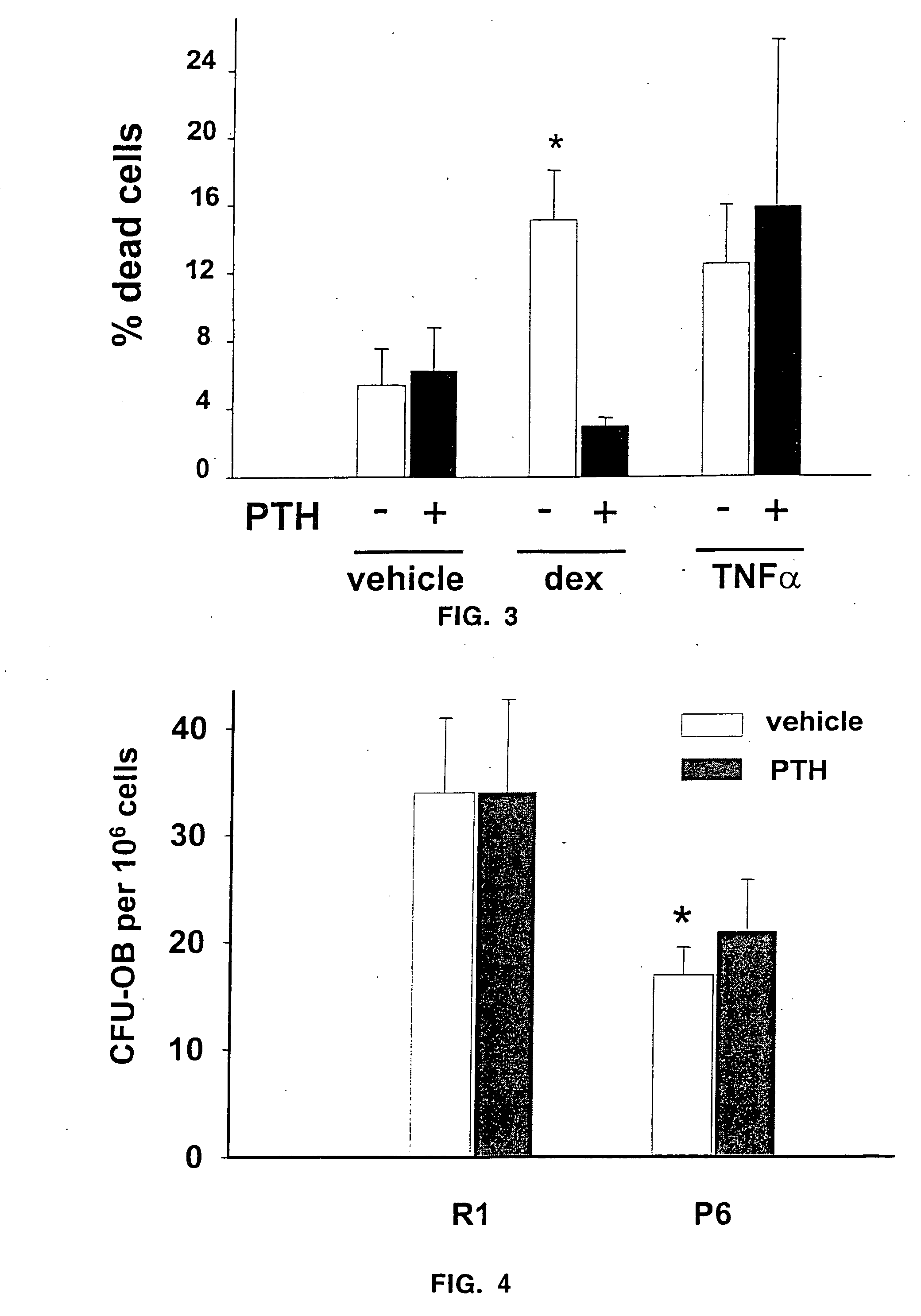
[0050] Osteoblastic cells were isolated from calvaria of 3- to 6-day-old C57 / B1 mice by sequential collagenase digestion. Cells were cultured for 5-8 ...

example 3

Mice

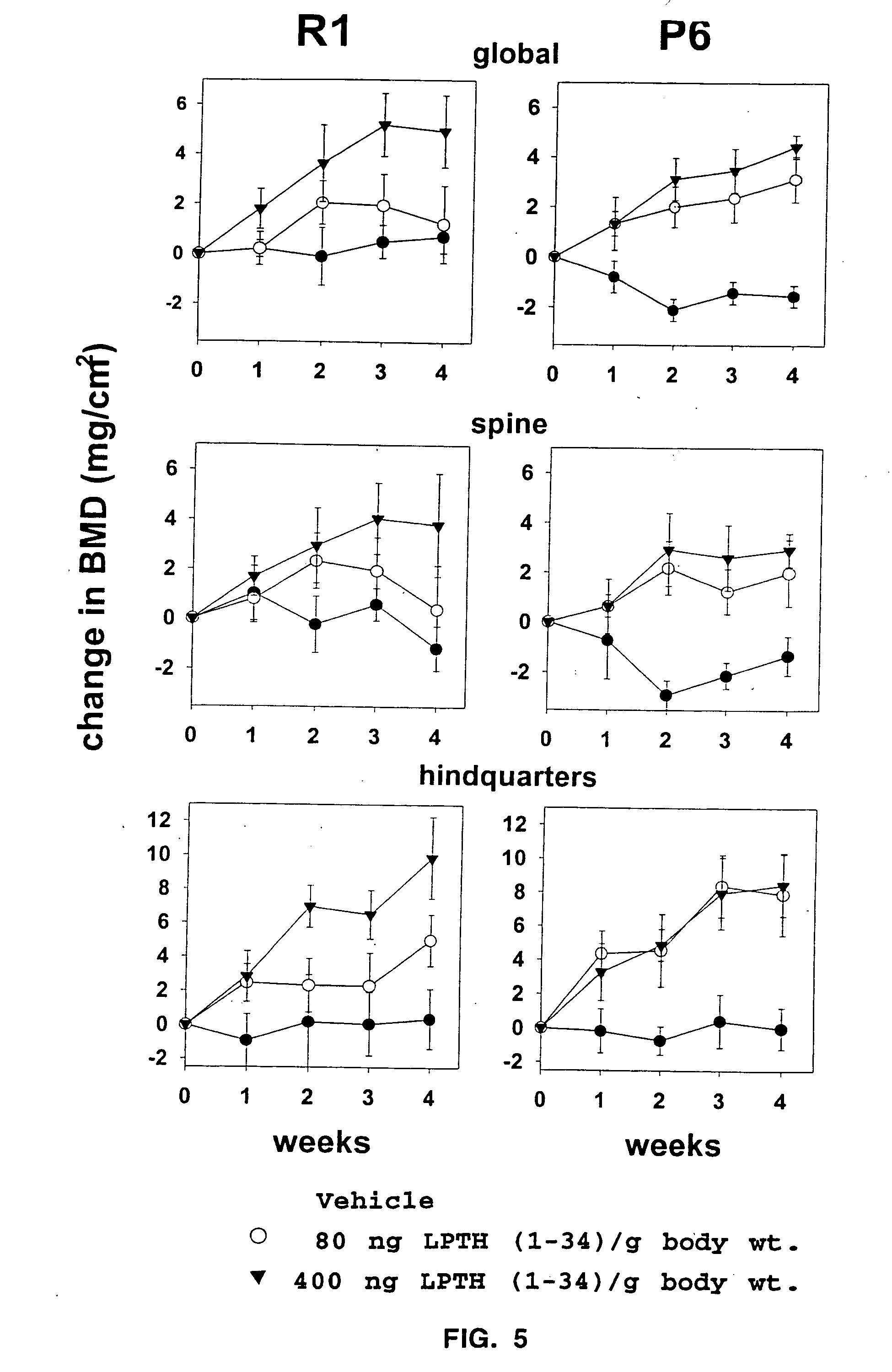
[0051] 4-5 month old male or female SAMR1 and SAMP6 were given daily injections of vehicle (0.9% saline, 0.01 mM β-mercaptoethanol, 0.1 mM acetic acid) or 400 ng / g body weight of hPTH(1-34) (Bachem, Torrence, Calif.) dissolved in vehicle (n=6-7 per group). Mice were fed a standard rodent diet (Agway RMH 3000, Arlington Heights, Ill.) ad libitum. The BMD of the spine and hindquarters was determined one day prior to initiation of the experiment (baseline scan) and at weekly intervals thereafter using dual-energy X-ray absorptiometry (QDR 2000, Hologic, Inc.) as described previously (3). The evaluation of each scan was based on the exact positioning and region of interest placement of the baseline scan using the “Compare” technique (4).


PUM

PropertyMeasurementUnit
molar concentrationsaaaaaaaaaa

Abstract

The present invention demonstrates that human parathyroid hormone 1-34 [hPTH(1-34)] exerts anti-apoptotic effects on osteoblasts when administered in an intermittent fashion to mice in vivo. The present invention further demonstrates that bovine PTH(1-34) [bPTH(1-34)] prevents glucocorticoid-induced apoptosis of osteoblastic and osteocytic cells in vitro. Therefore, the present invention demonstrates that the previously established anabolic effects of PTH on the skeleton are mediated by its ability to postpone osteoblast apoptosis, as opposed to a stimulatory effect on osteoblastogenesis. The present invention provides methods of screening agents for anti-apoptotic effects on osteoblasts, wherein such agents stimulate and / or restore bone in osteopenic individuals, or prevent bone loss caused by agents such as glucocorticoids.

Description

RELATED APPLICATIONS [0001] This application is a continuation of U.S. application Ser. No. 09 / 413,785, filed Oct. 7, 1999, which claims the benefit of U.S. Provisional Application No. 60 / 116,409, filed Jan. 19, 1999 and U.S. Provisional Application No. 60 / 103,385, filed Oct. 7, 1998. The entire teachings of the above application(s) are incorporated herein by reference. FEDERAL FUNDING LEGEND [0002] This invention was produced in part using funds obtained through grant PO1-AG13918 from the National Institutes of Health. Consequently, the federal government has certain rights in this invention.BACKGROUND OF THE INVENTION [0003] 1. Field of the Invention [0004] The present invention relates generally to bone physiology. More specifically, the present invention relates to inhibiting apoptosis of osteoblasts and osteocytes. [0005] 2. Description of the Related Art [0006] Remodeling of the human adult skeleton is carried out by teams of juxtaposed osteoclasts and osteoblasts. Osteoclasts...

Claims


Application Information

Patent Timeline
Patent Type & Authority Applications(United States)
IPC IPC(8): A61K31/00A61K31/05A61K31/11A61K31/565A61K31/567A61K38/29A61K45/06A61K49/00G01N33/50G01N33/74
CPCA61K31/00G01N2500/00A61K31/11A61K31/565A61K31/566A61K31/567A61K31/568A61K38/29A61K45/06A61K49/0008G01N33/5008G01N33/5044G01N33/743G01N2333/723A61K31/05A61P19/10
Inventor MANOLAGAS, STAVROS C.JILKA, ROBERT L.WEINSTEIN, ROBERT S.BELLIDO, TERESITA
Owner MANOLAGAS STAVROS C
Why Eureka
Try Eureka