Protocols for making hepatocytes from embryonic stem cells
- Summary
- Abstract
- Description
- Claims
- Application Information
AI Technical Summary
Benefits of technology
Problems solved by technology
Method used
Image
Examples
example 1
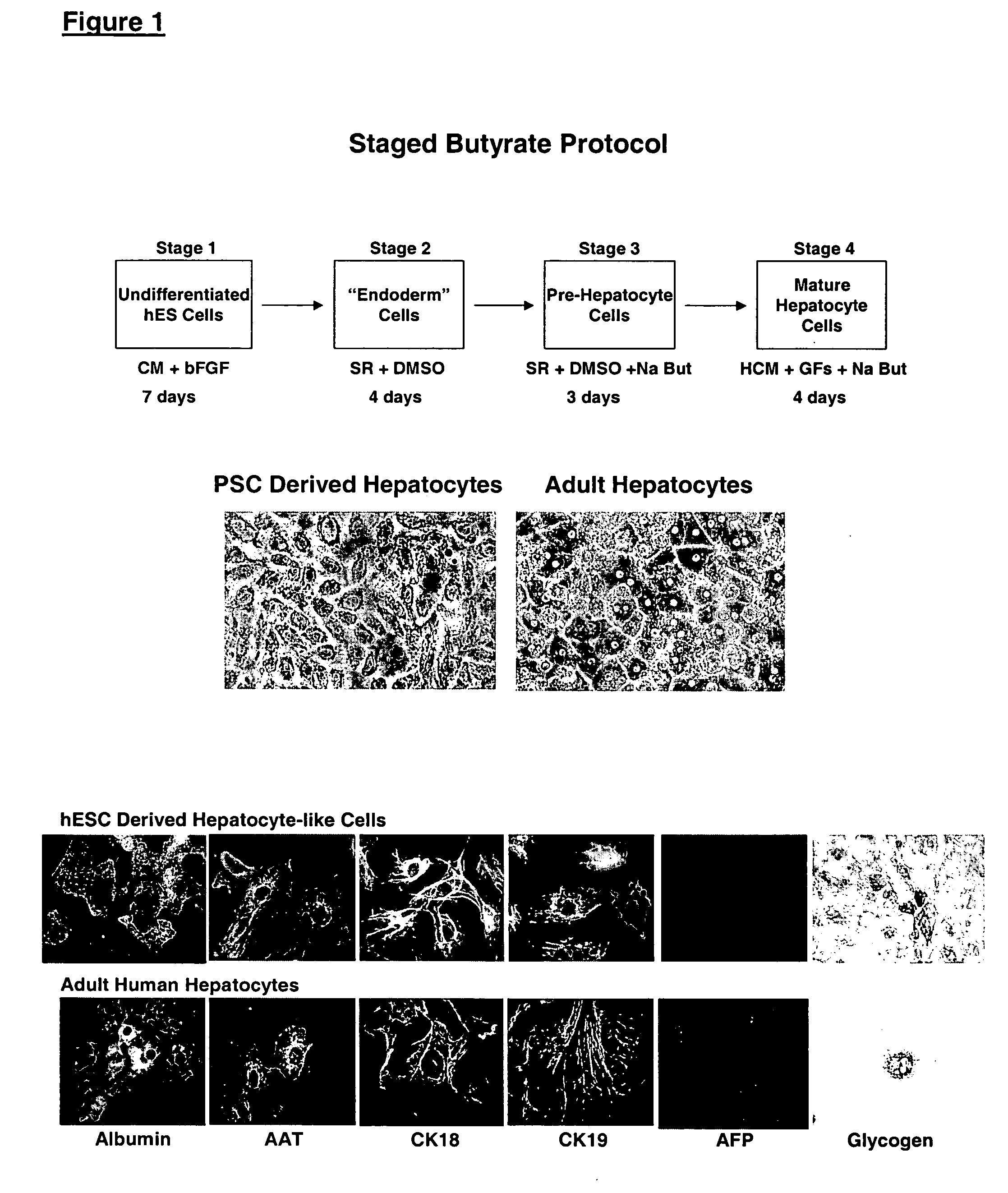
Differentiation of Human Embryonic Stem Cells Using n-butyrate
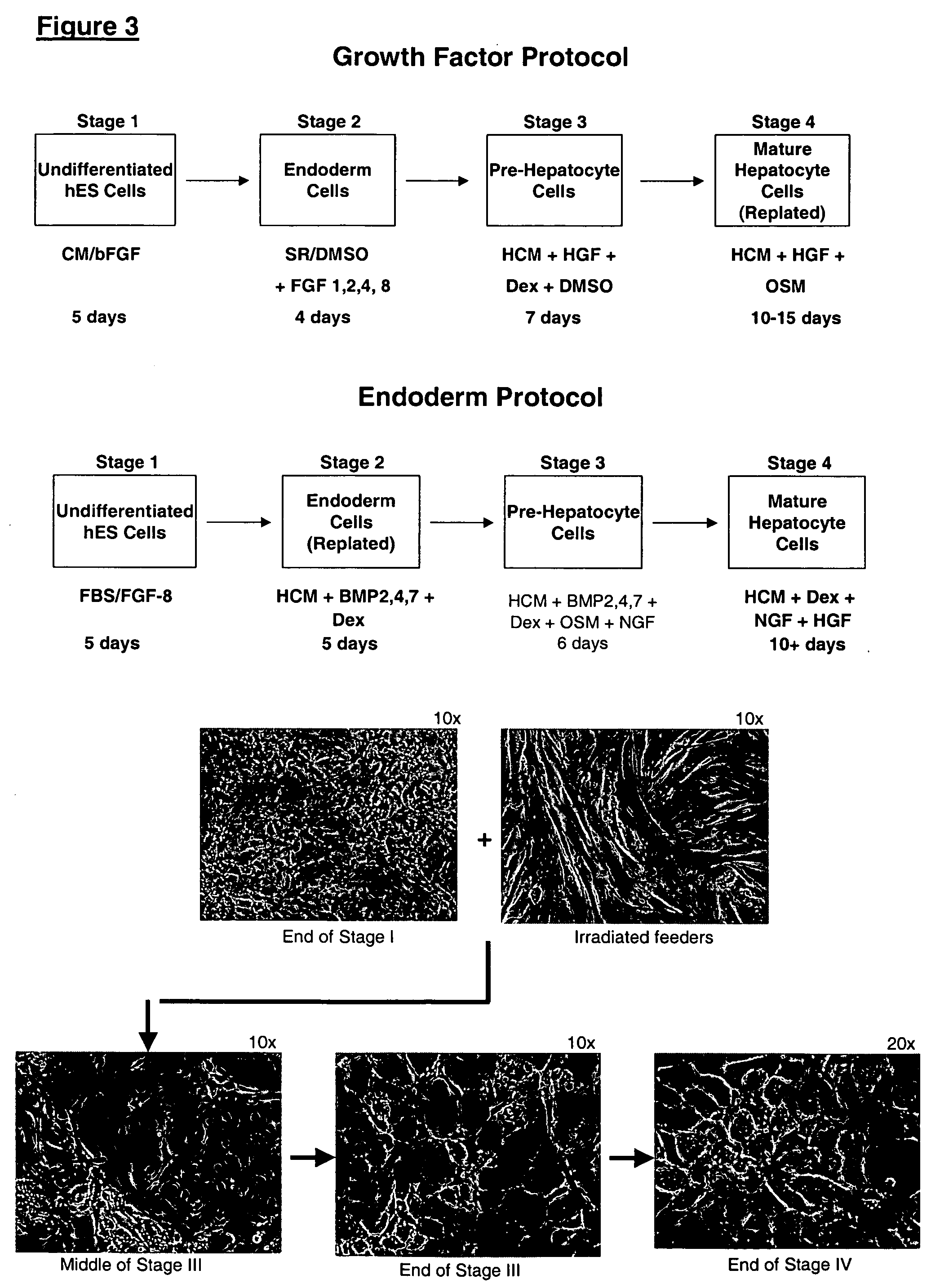
In this experiment, hES cells were maintained on primary mouse embryonic fibroblasts in serum-free medium according to standard methods. Embryoid bodies were formed by harvesting the cells with collagenase for 15-20 min, and plating dissociated clusters onto non-adherent cell culture plates (Costar) in a medium composed of 80% KO DMEM (Gibco) and 20% non-heat-inactivated FBS (Hyclone), supplemented with 1% non-essential amino acids, 1 mM glutamine, 0.1 mM β-mercaptoethanol.
The EBs were fed every other day. After 5 days in suspension culture, they were harvested and plated on Growth Factor Reduced Matrigel® coated plates and in chamber slides (Nunc). One of the following three conditions was used in parallel: medium containing 20% fetal bovine serum (FBS); medium containing 20% FBS and 5 mM sodium butyrate (Sigma); medium containing 20% FBS, 0.5% DMSO (ATCC), 4 μM dexamethazone (Sigma), 150 ng / mL insulin, 10 ng / mL E...
example 2
Marker Analysis of hES Cell Derived hepatocytes
hES derived embryoid bodies were plated on Matrigel® coated 6-well plates (for RNA extraction) and chamber slides (for immunocytochemistry) in medium containing 20% FBS and 5 mM sodium n-butyrate. The morphology of the differentiated cells was remarkably uniform, showing a large polygonal surface and binucleated center characteristic of mature hepatocytes.
On the sixth day after plating in the differentiation agent, the cells were analyzed for expression of markers by RT-PCR and immunocytochemistry. Glycogen content in these cells was determined using periodic acid Schiff stain. The number of cells in S phase of cell cycle was determined by incubating the cells with 10 μM BrdU on day 5 after plating, and subsequently staining with anti-BrdU antibody 24 hours later.
A summary of the phenotype analysis is provided in Table 4. Albumin expression was found in 55% of the cells. AFP was completely absent. Glycogen was being stored in at l...
example 3
Differentiation of hES to hepatocyte-Like Cells without Forming Embryoid Bodies
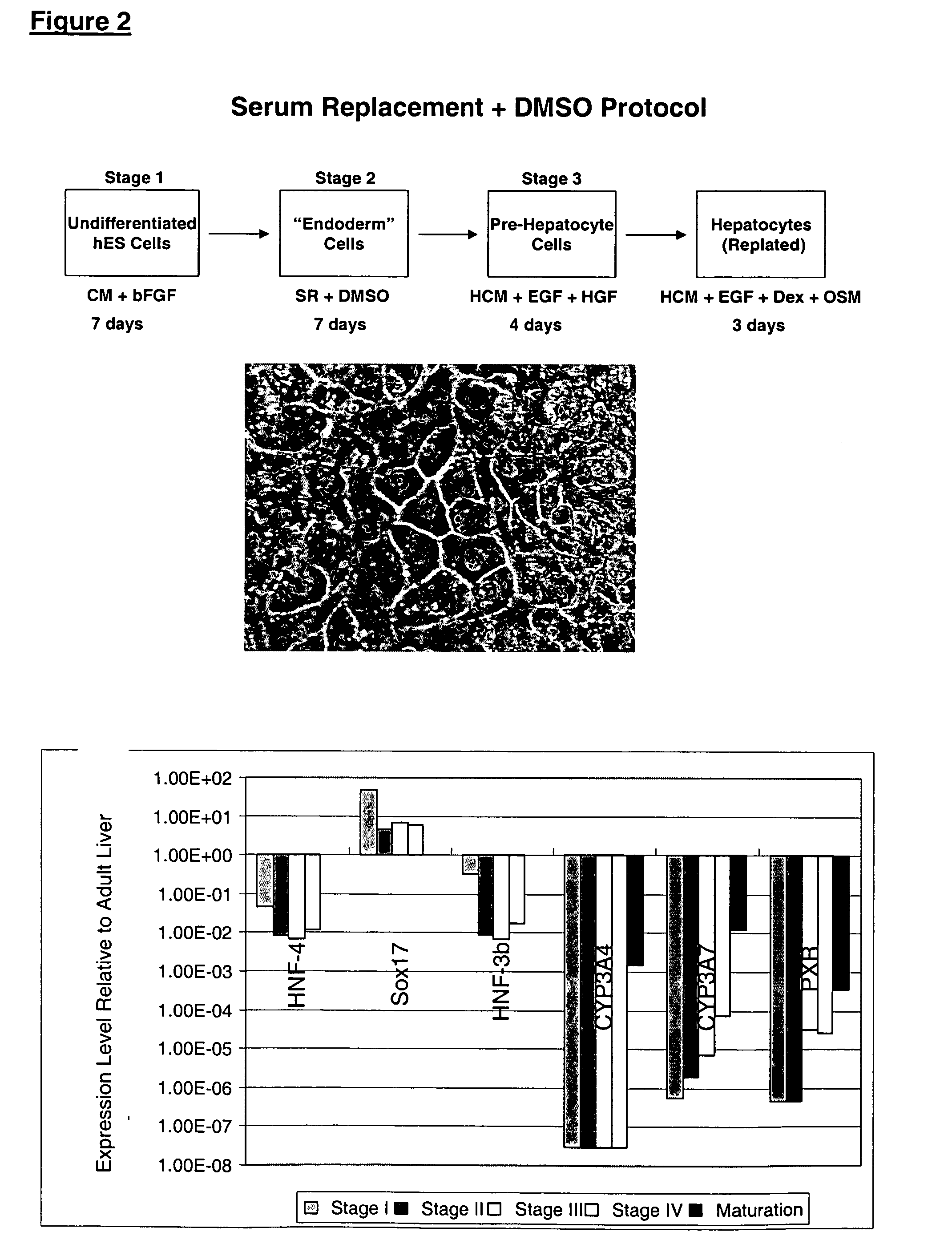
The undifferentiated hES cells were maintained in feeder-free conditions using medium conditioned by mouse embryonic fibroblasts, as previously described (WO 01 / 51616). The strategy was to initiate a global differentiation process by adding the hepatocyte maturation factors DMSO or retinoic acid (RA) to a subconfluent culture. The cells were then induced to form hepatocyte-like cells by the addition of Na-butyrate.
The hES cells were maintained in undifferentiated culture conditions for 2-3 days after splitting. At this time, the cells were 50-60% confluent and the medium was exchanged with unconditioned SR medium containing 1% DMSO. The cultures were fed daily with SR medium for 4 days and then exchanged into unconditioned SR medium containing 2.5% Na-butyrate. The cultures were fed daily with this medium for 6 days, at which time one half of the cultures were evaluated by immunocytochemistry. The oth...
PUM
 Abstract
Description
Claims
Application Information
 - R&D
- Intellectual Property
- Life Sciences
- Materials
- Tech Scout
- Unparalleled Data Quality
- Higher Quality Content
- 60% Fewer Hallucinations
Browse by: Latest US Patents, China's latest patents, Technical Efficacy Thesaurus, Application Domain, Technology Topic, Popular Technical Reports.
© 2025 PatSnap. All rights reserved.Legal|Privacy policy|Modern Slavery Act Transparency Statement|Sitemap|About US| Contact US: help@patsnap.com


