
X-ray examination method and apparatus with automatic gating of the x-ray beam

a technology of x-ray beam and x-ray examination method, which is applied in the direction of instruments, diaphragms/collimeters, diaphragms for radiation diagnostics, etc., can solve the problems of increasing the time-consuming and laborious of manual adjustment of collimators, and reducing radiation exposure. , to achieve the effect of reducing radiation exposur

Inactive Publication Date: 2005-01-20
SIEMENS AG
View PDF5 Cites 21 Cited by
  • Summary
  • Abstract
  • Description
  • Claims
  • Application Information

AI Technical Summary

Benefits of technology

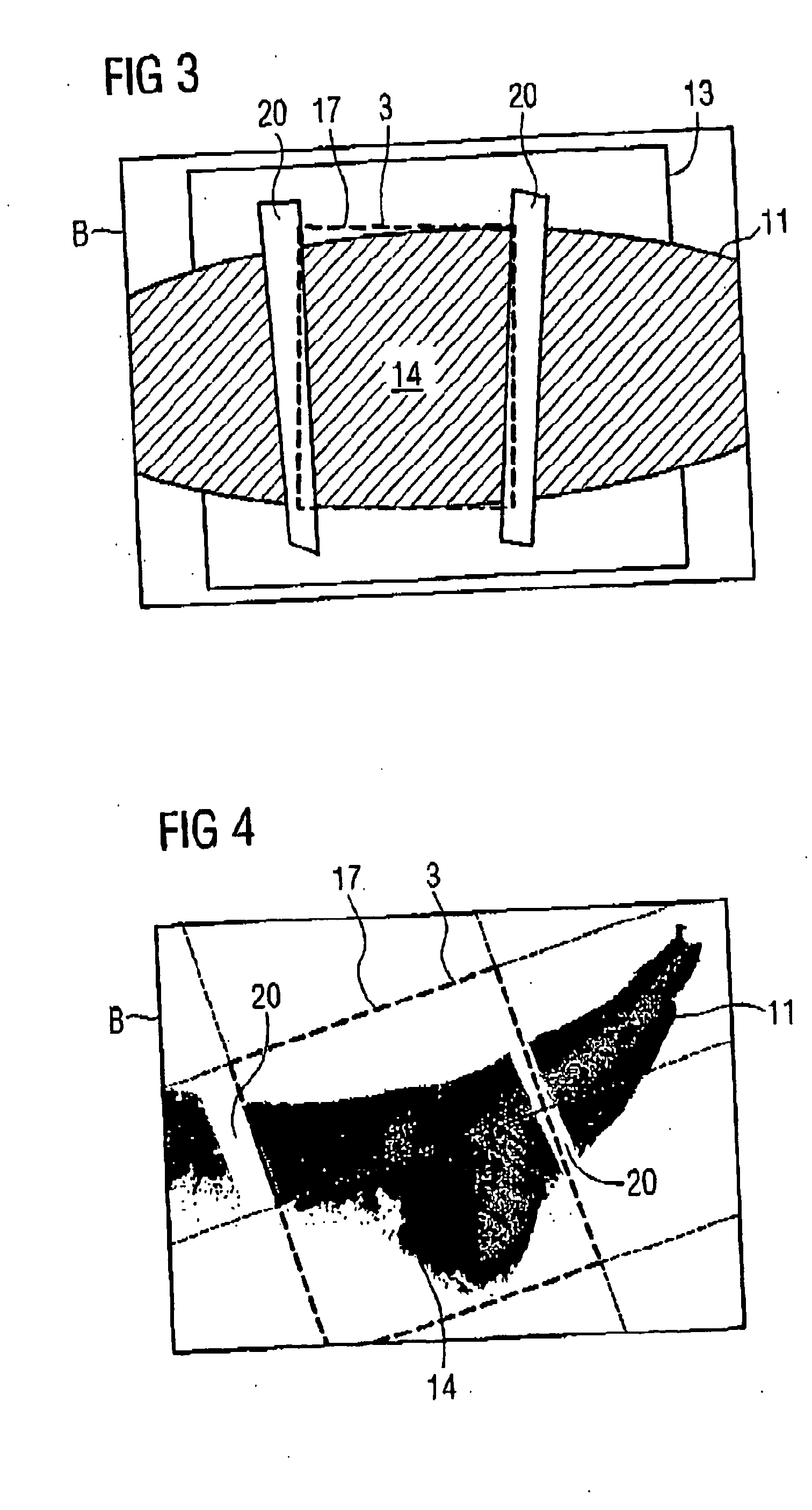
[0013] The selection of the area to be exposed ensues essentially automatically via the evaluation unit. This automation is particularly facilitated because the area is not determined using the actual x-ray apparatus, but instead is determined virtually using the acquired adjustment image. As a result of the automation, the collimation ensues extremely quickly, meaning without noticeable time loss. The x-ray acquisition therefore can be started as soon as the subject to be examined is positioned. Furthermore, a particularly precise setting of the area techniques, rather than by the inspection of a technician. The radiation exposure for the patient thus can be kept particularly low.
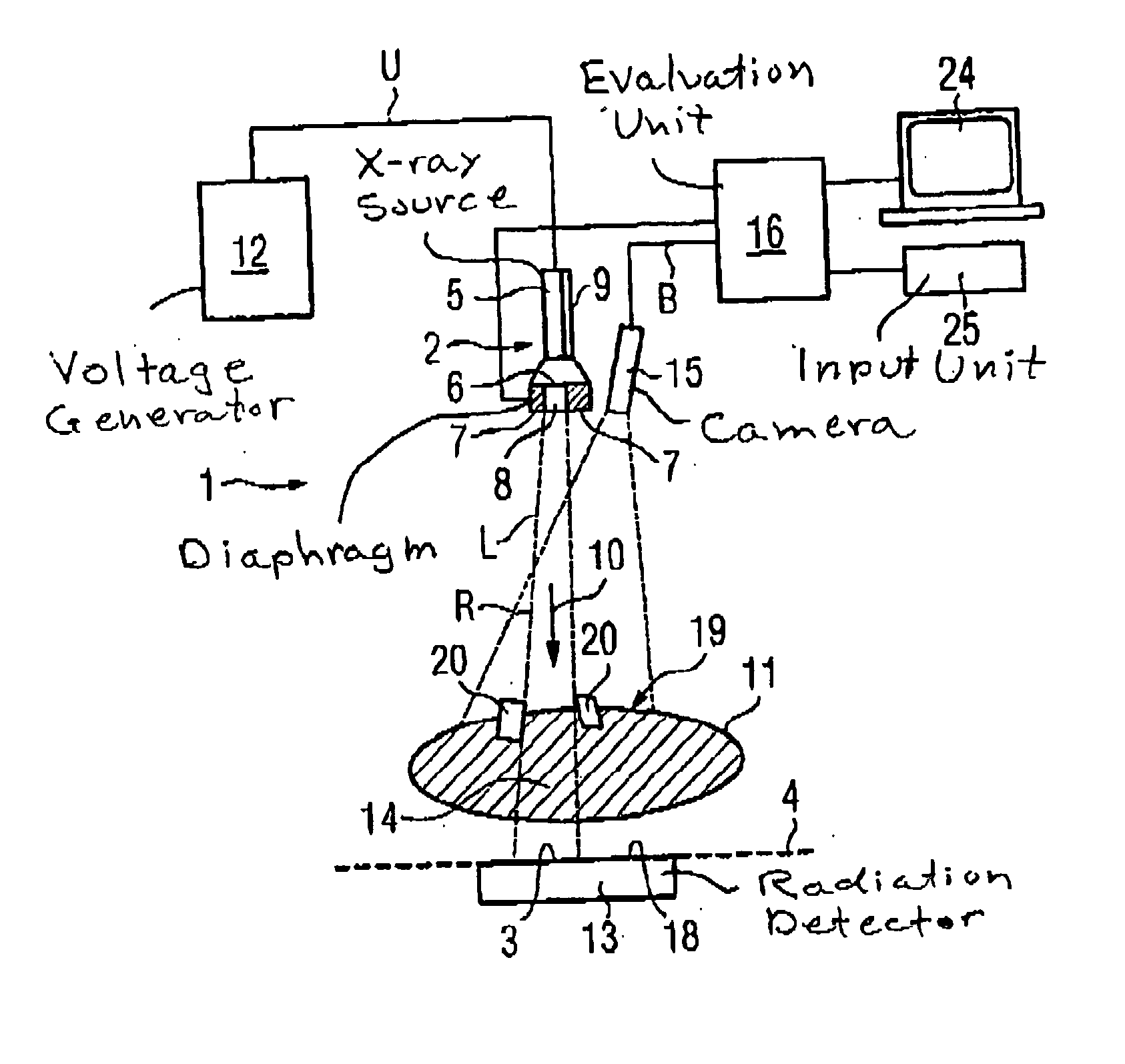
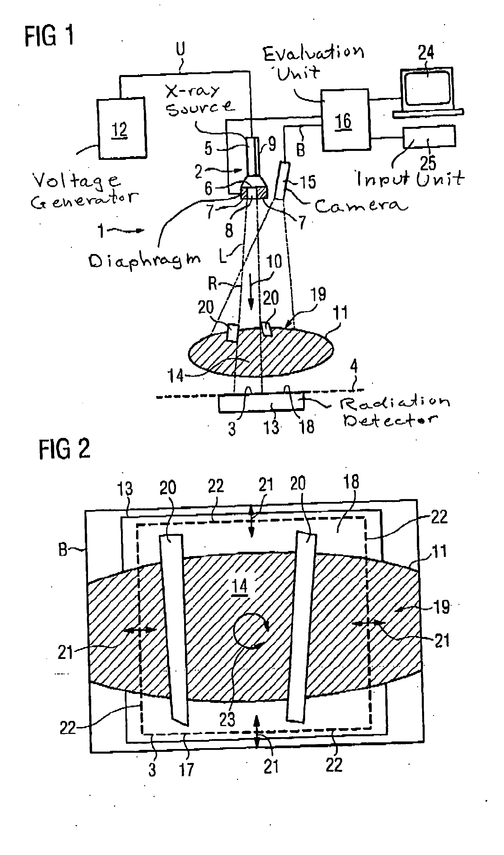
[0020] A light source preferable is operated in parallel with the x-ray radiator in order to visualize the area section adjusted by the diaphragm for the technician. The open diaphragm region (meaning the region of the diaphragm permeable to visible light and x-ray radiation) is projected on the x-ray exposure plane by the visible light generated by the light source, so the technician can monitor in a simple manner whether the area to be exposed has been correctly set in the aforementioned automated procedure. In order to manually readjust the area as necessary, the x-ray apparatus preferable also allows for manual adjustment of the diaphragm. This can ensue by data input and output units connected to the evaluation unit, such as a monitor, keyboard, mouse, etc., and operating software integrated into the evaluation unit.
[0021] If a number of modifications of the area are selectable in which the region of the subject to be examined is identically inscribed, the area thereby optimized in the course of the selection process, to the effect that its surface content is minimal. This surface minimization, which serves to reduce the radiation exposure, preferably is implemented with a mathematical minimization technique.

Problems solved by technology

The adjustment process, which is subsequently also designated as a “collimation”, is relatively time-consuming.
This is disadvantageous insofar as the patient must remain substantially motionless during the gating and the subsequent exposure.
Using conventional technology, these requirements run counter to one another, particularly because manual adjustment of the collimator normally becomes increasingly more time-consuming the more precisely the area to be exposed is matched to the region to be examined.

Method used


Image

  • X-ray examination method and apparatus with automatic gating of the x-ray beam
  • X-ray examination method and apparatus with automatic gating of the x-ray beam
  • X-ray examination method and apparatus with automatic gating of the x-ray beam

Examples


Embodiment Construction

[0028] The x-ray apparatus 1 shows in FIG. 1 has an exposure unit 2, by means of which a predeterminable area 3 of an x-ray exposure plane 4 can be exposed with x-ray radiation R.

[0029] The exposure unit 2 includes an x-ray radiator 5 to generate the x-ray radiation R as well as a diaphragm 6. The term “diaphragm” In radiology means a device that includes a material (for example, lead) that significantly absorbs x-ray radiation R and that serves to gate or focus beams as well as to shield from scattered radiation (see, for example, “Psychrembel Klinisches Wöterbuch”, 259th new edited edition. Berlin: De Gruyter, 2002, P. 879). The diaphragm 6 according to FIG. 1 has a number of adjustable plates or lamellae 7 that delimit a radiation-permeable region 8. The x-ray radiation R penetrating through this region 8 is incident within the area 3 on the x-ray exposure plane 4. In contrast, the region of the x-ray exposure plane 4 lying outside of the area 3 is shadowed from the x-ray radiat...


PUM

PropertyMeasurementUnit
areaaaaaaaaaaa
coloraaaaaaaaaa
area contentaaaaaaaaaa

Abstract

An x-ray apparatus has an exposure unit with an x-ray radiator and a diaphragm in front thereof, and a camera system with which an adjustment image is acquired of a subject positioned in front of an x-ray exposure plane. Using the adjustment image, a region to be examined of the subject is identified and an area of the x-ray exposure plane is selected such that the projection of the region to be examined of the subject is substantially inscribed in the projection of the areal section. The diaphragm is adjusted such that the x-ray radiation generated by the x-ray radiator is incident on the x-ray exposure plane only within the selected areal section.

Description

BACKGROUND OF THE INVENTION [0001] 1. Field of the Invention [0002] The present invention concerns an x-ray examination method as well as an x-ray apparatus to implement the method. [0003] 2. Description of the Prior Art [0004] Particularly in medical technology) imaging examination methods based on the acquisition of x-ray Images have increased for years. In general, the x-rays generated by an x-ray radiator are emitted in the direction of an x-ray exposure plane in which the x-ray radiation is detected. Classically, photographic x-ray films are used for detection of the x-ray radiation. More recently digital x-ray detectors also have been used as an alternative. [0005] For the acquisition of the x-ray image, a subject to be examined, which for medical purposes normally is a body part of a patient, is positioned in the beam path of the x-ray radiation in front of the x-ray exposure plane. The image contrast results due to the spatially different strong attenuation of the x-rays pen...

Claims


Application Information

Patent Timeline
Patent Type & Authority Applications(United States)
IPC IPC(8): A61B6/06A61B6/08G21K1/04
CPCA61B6/06G21K1/04A61B6/08
Inventor HORNEGGER, JOACHIM
Owner SIEMENS AG
Learn More