
Controlled enzymatic removal and retrieval of cells

Inactive Publication Date: 2004-09-23
RAMOT AT TEL AVIV UNIV LTD
View PDF28 Cites 21 Cited by
  • Summary
  • Abstract
  • Description
  • Claims
  • Application Information

AI Technical Summary

Benefits of technology

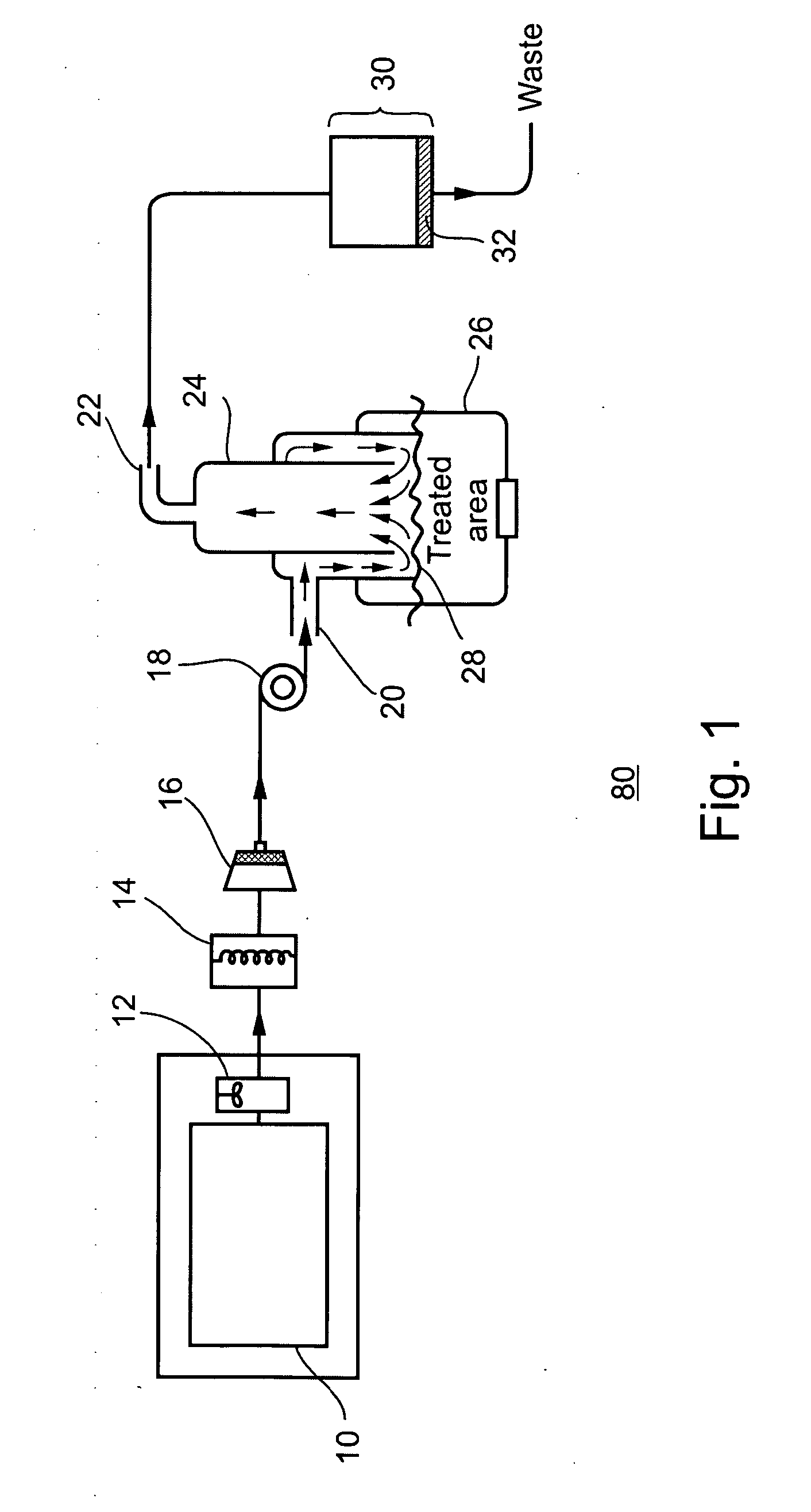
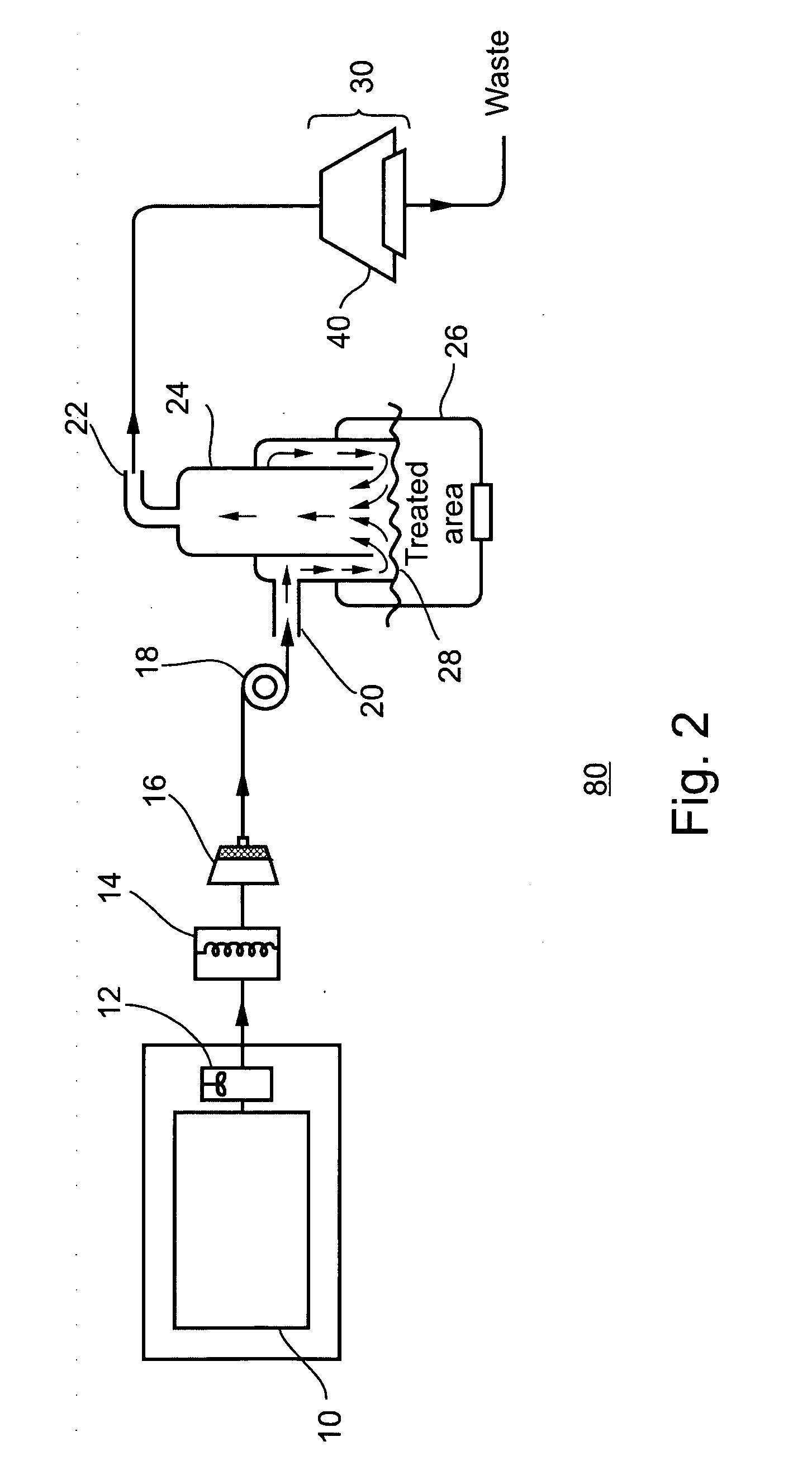
[0050] According to another aspect of the present invention, there is provided a device for removing cells from a skin portion of a subject, comprising: (a) a first reservoir containing an effective amount of at least one protease in a non-aqueous catalytically inactive form; (b) a first receptacle for receiving the first reservoir; (c) a second reservoir containing a protease activating solution, the activating solution activates catalytic activity of the at least one protease upon mixing with the at least one protease; (d) a second receptacle for receiving the second reservoir; (e) a mixing mechanism in fluid communication with the first and second reservoirs when received by the first and second receptacles, the mixing mechanism is for mixing the at least one protease and the activating solution such that the at least one protease becomes catalytically active in solution; (f) an applicator in fluid communication with the mixing mechanism, for restricting streaming of the active protease solution, over, and in contact with, the skin portion, the applicator includes: (i) an inlet port for receiving the active protease solution from the mixing mechanism; (ii) a first tube structure operatively connected to the inlet port for directing the streaming of the active protease solution from the inlet port to a treatment zone of the skin portion, such that the streaming active protease solution enzymatically and mechanically causes the removal of cells from the skin portion; (iii) a second tube structure positioned within the first tube structure for adjustably directing the streaming active protease solution and the removed cells away from the treatment zone, wherein a screw mechanism operatively connected to the second tube structure allows adjustment of height of opening of the second tube structure with respect to a -skin-facing opening of the treatment zone; and (iv) an outlet port operatively connected to the second tube structure for removing the streaming active protease solution and the removed cells from the second tube structure, thereby removing the cells from the skin portion of the subject.

Problems solved by technology

Surgical procedures are generally painful and destructive to the healthy, neighboring tissues, resulting in scarring and abnormal pigmentation of the treated areas, for example, as described by Gambichler, T., Senger, E., Rapp, S., Almouti, D., Altmeyer, P., and Hoffman, K., in "Deep Shave Excision Of Macular Melanocyte Nevi", Dermatol. Surg., Jul. 26 (7), 662-66 (2000), the need for administration of anesthesia, and significant stress trauma to the patient, for example, as described by Augustin, M., Zschocke, I., Godau, N., Buke-Kirschenbaum, A., Peschen, M., Sommer, B., and Sattler, G., in "Skin Surgery Under Local Anesthesia Leads To Stress-induced Alterations Of Psychological, Physical And Immnune Functions", Dermat. Surg., November 25 (11), 868-71 (1999).
In addition, surgical excision of certain lesions is often complicated by the presence of more than one aberrant cell type, for example, as described by Crawford, J. B., Howes, E. L. Jr., and Char, D. A., in "Conjunctival Combined Nevi", Trans-Am. Ophthamol. Soc., 97, 170-83 (1999), and is inappropriate for sensitive and precarious anatomical regions.
These disadvantages of surgical excision of skin lesions have led to the development of non-mechanical methods such as electro-cauterization, electro-ablation, cryosurgery with liquid nitrogen and lasers.
However, there remain the problems of pain and scarring associated with the intense heat required for tissue ablation, poor results with attempts at laser treatment of certain lesions, such as melanocytic and congenital nevi (Raulin, C. et al., same as above) and the importance of removal, rather than ablation of the abnormal tissue for histological analysis to determine the character and extent of the lesion.
However, no mention is made of control of enzyme activity once applied, or of a means of obtaining cells from the treated tissues.
However, the procedure described relates to preparation of cells for tissue culture from tissue fragments rather than the therapeutic application of cell removal from live tissue.
Furthermore, no provision is made for collection of cells from living tissue in situ
Proteolytic enzymes, being proteins, are in themselves substrates for self-digestion, or autolysis, thus limiting the effectiveness of active, protease based preparations.
Enzyme stability is enhanced when dry, but solubility and even dispersal of the enzyme solid in the activating buffer is difficult, resulting in poor control of enzyme activity at point of delivery.
However, the methods of treatment with these enzymes have been limited to topical application and intradermal injection.
In addition to the discomfort and protracted character of such a treatment regimen, no retention of cells from the lesions is made possible, necessitating conventional, surgical biopsy methods prior to enzymatic treatment.
Thus, cell collection or retention from the treated area is not possible and, as in other dermatological enzyme treatment protocols, no control of protease activity after administration is afforded.
Topical application, or injection of proteases offers little control over the level of catalytic activity remaining in situ, with autoproteolytic and normal dermal lytic and acidic processes causing unpredictable degradation.
Conventional mechanical and non-mechanical methods of treating and removal of skin lesions such as razor-blade or scalpel excision, CO.sub.2 laser surgery, cryosurgery, electrocauterization, and electroablation are associated with pain, stress trauma, bleeding, scarring, contamination, hyperpigmentation and disruption of adjacent and underlying tissue.
Active shelf life of the protease is limited, and precise control of enzyme activity at the site of administration is virtually unattainable, once injection or topical application is completed.
None of the aforementioned examples, however, relate to the administration of proteases for treatment of living cells, nor provide for ongoing, precise control of the activation of catalytic activity at the site of application.
However, powdered, lyophilized or granulated enzyme preparations are often difficult to disperse homogeneously in diluent solutions.
Treatment without determining accurate diagnosis may remove the lesion, but will often incur unnecessary scarring, recurrences, and financial hardships.
As mentioned above, previous methods of non-surgical treatment of skin lesions, such as laser surgery, electrosurgery and chemical or enzymatic ablation have not provided any means for obtaining cells from the lesions, necessitating the use of traditional surgical biopsy techniques for accurate diagnosis.

Method used


Image

  • Controlled enzymatic removal and retrieval of cells
  • Controlled enzymatic removal and retrieval of cells
  • Controlled enzymatic removal and retrieval of cells

Examples


Embodiment Construction

[0144] Enzymatic Epidermis Removal

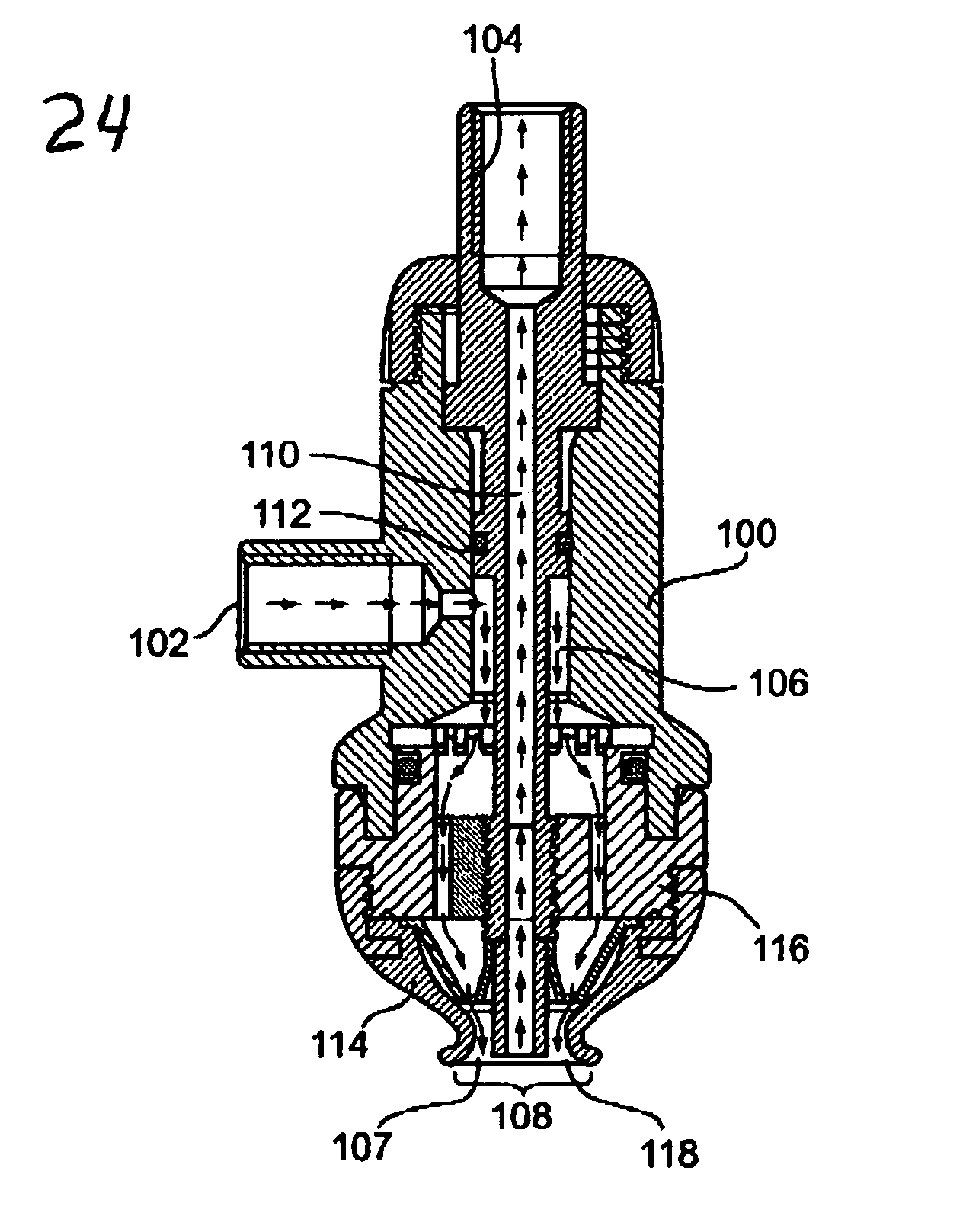
[0145] Design and fabrication of an applicator for the treatment of skin surface area 9 mm in diameter: An applicator for the treatment of skin areas, 9 mm in diameter, was configured as illustrated in FIG. 10. The applicator was fabricated in a workshop with special emphasis on making the skin contacting surface from silicone to ensure flexibility and effective sealing.

[0146] Briefly, reference is now made to FIG. 10, a cross sectional view of an exemplary specific preferred embodiment of the applicator included in the device of the present invention, which is applicable for practicing the present invention on skin. Applicator 24 used in this experiment includes a housing 100 having an inlet 102 and an outlet 104. Fluid entering through inlet 102 is directed via a first tube structure 106 to a treatment zone 107 defined by a somewhat conical silicon structure 114 having a skin-facing opening 108, 9 mm in diameter. A second tube stature 110 position...


PUM

PropertyMeasurementUnit
Temperatureaaaaaaaaaa
Structureaaaaaaaaaa
Catalytic activityaaaaaaaaaa

Abstract

Method and device, including a uniquely operative applicator, and pharmaceutical compositions, for the controlled, non-surgical removal and retrieval of cells from a variety of skin lesions and tissue surfaces are disclosed. A synergistic effect of proteolytic digestion of the intracellular matrix and "stripping" flow is achieved by treating a defined area with a controlled, continuous stream of proteolytic enzyme solution, causing gentle but effective tissue erosion. Isolated cells from the skin lesion and / or tissue surface may be collected from the protease solution stream for histological analysis and / or cell culture, affording a method of "enzymatic biopsy". The protease solution may be supplemented with anesthetics, coagulants, anticoagulants and antibiotics to decrease the discomfort, erythema, bleeding, risk of infection and scarring traditionally associated with surgical treatment of skin lesions. Delivery of precise levels of catalytic activity is ensured by controlled activation of stable, inactivated enzyme stock solutions and powders shortly prior to application.

Description

[0001] This is a continuation-in-part of PCT international patent application No. IL02 / 00572, filed Jul. 16, 2002, entitled: "Device For And Method Of Controlled Enzymatic Removal And Retrieval Of Tissue", claiming priority from U.S. patent application Ser. No. 09 / 915,518 filed Jul. 27, 2001, of same title; both specifications of which are incorporated herein by reference.FIELD AND BACKGROUND OF THE INVENTION[0002] The present invention relates to a method, device, and pharmaceutical compositions, for the controlled removal of cells from the surface of viable tissue by continuous local application of a solution containing a proteolytic enzyme and, more particularly, to a method, device and pharmaceutical composition for non-surgical, enzymatic treatment and biopsy of skin lesions. The method and device each includes a uniquely configured and operative applicator including at least one inlet and at least one outlet, each providing a passageway for streaming of a solution therethrough...

Claims


Application Information

Patent Timeline
IPC IPC(8): A61B10/00A61B10/02A61B17/00A61B17/22A61B17/34A61B17/54A61K38/48A61K38/49A61M3/02A61M27/00A61M35/00C12N5/071
CPCA61B10/0035C12N2509/00A61B17/545A61B2017/00761A61B2017/00769A61B2017/00774A61B2017/22082A61B2017/3411A61K38/48A61K38/4886A61M1/0088A61M3/0241A61M3/0258A61M3/0283A61M3/0287A61M27/00C12M45/09C12N5/0629A61B10/02A61M3/0204A61M1/77A61M35/30A61K38/4813A61K38/4873A61K38/488A61B2010/0225A61M1/90
Inventor FREEMAN, AMIHAY
Owner RAMOT AT TEL AVIV UNIV LTD
Learn More