
Method and device for electroporation of biological cells

a biological cell and electroporation technology, applied in the direction of biomass after-treatment, enzymology, specific use bioreactors/fermenters, etc., can solve the problems of reducing the survivability of cells, reducing the number of available cells, and requiring careful and time-consuming optimisation of electropermeabilisation protocols

Inactive Publication Date: 2003-08-07
EPPENDORF AG
View PDF3 Cites 17 Cited by
  • Summary
  • Abstract
  • Description
  • Claims
  • Application Information

AI Technical Summary

Benefits of technology

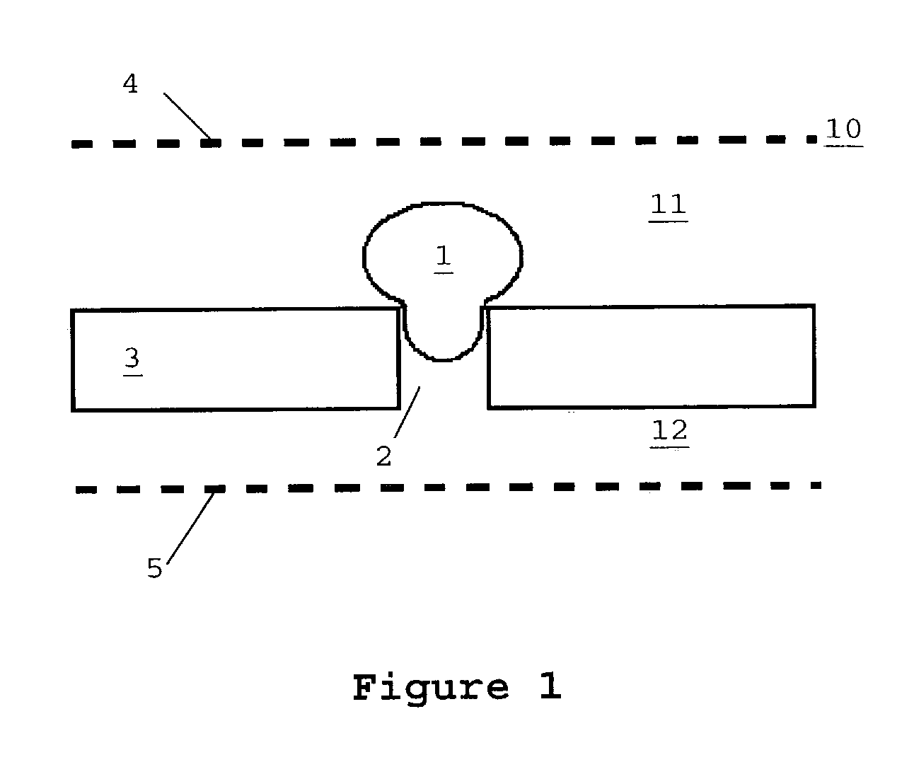
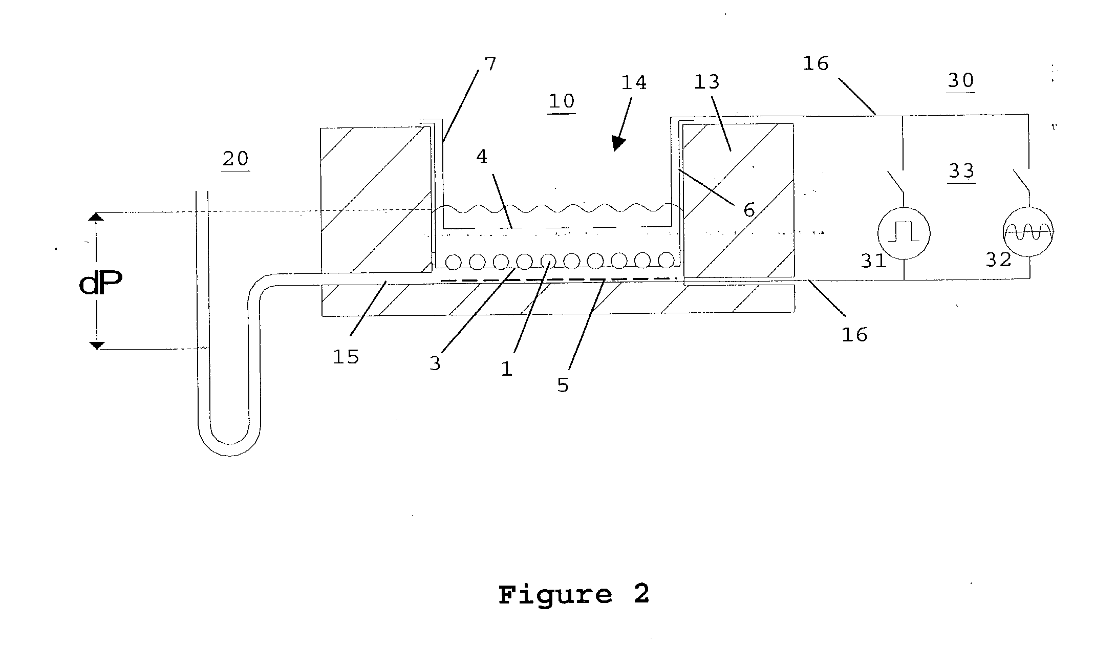
[0019] In contrast to conventional electropermeabilisation methods which use very high field intensities and input voltages, if need be, the method according to the invention works with middle or even low input voltages. This is possible because, as a result of the arrangement according to the invention, of the carrier element, the applied field is forced to flow through the cells embedded in the pores. This encourages charging and break-through of the cell membrane. Furthermore, the low hydrostatic negative pressure to which the cells are exposed causes deformation of the cells in the direction of the field. Advantageously, as illustrated below, this additional mechanical stress results in further improvement of the electropermeabilisation yield. A further advantage of the invention consists of electropermeabilisation being able to be carried out in physiological media, i.e. in media rich in salt. In contrast, conventional methods for electropermeabilisation of suspension cells are carried out predominantly in media of low conductivity, because permeabilisation in such media with a lower ion content takes place more efficiently. By using physiological media in combination with the mechanical support provided by the carrier element, the survivability of the cells after electropermeabilisation is increased significantly, without having a significant influence on the electropermeabilisation yield.

Problems solved by technology

However, both very strong electrical fields and media of low conductivity reduce survivability of the cells.
In the case of rare or valuable cells (for example genetically modified hybridoma cells or dendritic cells) this can in turn significantly reduce the number of available cells.
For this reason, very careful and time-consuming optimisations of the electropermeabilisation protocols are necessary for suspension cells.
However, in the case of electroporation of suspended cells, this method is limited to cells of sufficient size (e.g. giant algae, xenopus oozytes) and cannot be used on most cells of animal or plant origin which are only a few micrometers in size.
While this technique makes possible electroporation and electrofusion in solutions of high or low conductivity, it has, however, the disadvantage in that the arrangement of the cells on the carrier element and the result of electrical treatment cannot be observed or monitored.
The device known from WO 93 / 02178 is thus unsuitable for the treatment, in particular, of rare or valuable cells under practical conditions.
The known device has a further disadvantage in that the chamber is combined with a closed pressure system.
The design of the device is expensive and handling in practical application is complicated.
While the system described by Y. Huang et al. allows real-time monitoring of the poration process, it is, however, not suitable for practical applications in which a multitude of biological cells are to be treated.
Furthermore, direct current measuring only provides an information about the membrane characteristics during electroporation, without allowing any further-reaching characterisation of the treated cell for any subsequent processing steps.
However, this information is insufficient for practical electroporation applications, for example in the field of medicine.
Optical observation, however, only produces qualitative assessments.
Generally, there is a problem in that only the current is measured in conventional techniques.
There is no specification of measuring parameters that furnish reliable measuring results.
However, the measuring object comprising electrolyte and biological cell in the measuring chamber is a complicated structure characterised by capacities and resistances which, depending on the type of cells to be treated, requires other measuring parameters.

Method used


Image

  • Method and device for electroporation of biological cells
  • Method and device for electroporation of biological cells
  • Method and device for electroporation of biological cells

Examples


Embodiment Construction

[0037] Cells

[0038] The mouse myeloma cell line Sp2 was cultivated in the RMPI 1640 Complete Growth Medium (CGM) with 10% FCS (Fetal Calf Serum, PAA, Linz, Austria) at 37.degree. C. under 5% CO2. The cells were kept in the exponential growth phase by subcultivation three times per week. Prior to commencement of electropermeabilisation, the cells were washed once or twice in pulse medium and resuspended in the pulse medium 10 min prior to pulse application. The average cell diameter was determined by electronic size determination by means of CASY (Scharfe Systems, Reutlingen, Germany) at approx. 14 .mu.m.

[0039] Pulse Media

[0040] A phosphate buffer comprising 1.15 mM K2HPO4 / KH2PO4 buffer, pH 7.2 was used as a pulse medium. KCl at a concentration of 10 and 30 mM respectively was added as a conducting salt. Osmolarity was set to 290 mOsm by adding Inositol, so as to obtain an isoosmolar solution.

[0041] Impedance Measurement Prior to Electroporation

[0042] FIG. 4A shows impedance spectra o...


PUM

PropertyMeasurementUnit
Pore sizeaaaaaaaaaa
Translucencyaaaaaaaaaa
Frequencyaaaaaaaaaa

Abstract

Described are methods for electrical treatment of biological cells, in particular using electrical field pulses, involving the steps: arrangement of the cells (1) on apertures (2) of a solid planar carrier element (3) which divides a measuring chamber (10) into two compartments (11, 12) ; and temporary formation of an electrical treatment field which permeates the cells, wherein an alternating-current impedance measurement takes place on the carrier element (3), and from the result of the alternating-current impedance measurement, a degree of coverage of the carrier element and / or healing of the cells after electrical treatment are / is acquired. Also described are devices for implementing the methods.

Description

[0001] The invention relates to methods for electrical treatment of biological cells, in particular for electroporation or electropermeabilisation of biological cells which are arranged on a fixed carrier element, as well as electroporation devices for carrying out such methods.[0002] The technique of electroporation (also known as electropermeabilisation, electroinjection or electro-transfection) represents a commonly known means for the inward transfer of membrane-impermeable xenomolecules (e.g. dyes, drugs, hormones, proteins, plasmids etc.) into live cells, or for the controlled release of intracellular substances from the cells. Electroporation has become widely used and recognised because it is more controllable, more reproducible and more efficient than other (chemical or viral) methods of intracellular transport of foreign molecules. The technique of electroporation is based on a temporary increase in membrane permeability which is caused by a reversible electrical break-thr...

Claims


Application Information

Patent Timeline
IPC IPC(8): C12M1/42C12N13/00
CPCC12N13/00C12M35/02
Inventor ZIMMERMANN, ULRICHSUKHORUKOV, VLADIMIRLUCAS, KURT
Owner EPPENDORF AG
Learn More