
Highly cost-effective analytical device for performing immunoassays with ultra high sensitivity

Inactive Publication Date: 2002-08-15
COUNCIL OF SCI & IND RES +1
View PDF0 Cites 39 Cited by
  • Summary
  • Abstract
  • Description
  • Claims
  • Application Information

AI Technical Summary

Benefits of technology

[0048] The main object of the present invention is to provide an improved highly cost-effective, easy to manufacture analytical device based on Flow-through principle for performing rapid immunoassays for visual detection of diagnostically important antigen or antibody.
[0049] Another object of the invention is to use disposable absorbent body for not only making construction of the device simpler but also for application of elements of signal amplification for attaining ultra high sensitivity.
[0050] In yet another object of the invention is to develop a simple assay method with the invented device which without spreading allows always downward flow of sample or costly labeled reagents efficiently, without application of any force to the absorbent body thereby overcoming above described drawbacks of the hitherto known Flow-through devices.Novelty

Problems solved by technology

These antigen or antibodies are present in biological fluid such as serum in extremely minute amounts and early detection sometimes is not possible.
1. The detection limit of the method with different substrate solution depending on the enzyme label used for streptavidin is in the range of 80 to 1 pg.
2. The increased in sensitivity is only 30-fold.
3. Long incubation periods required makes the method time consuming.
As the membranes are very delicate and difficult to handle, numerous assay devices, in various configurations were developed for wider use under field conditions.
1. Method is time consuming and often requires a number of manipulative steps, for example, the addition and incubation of assay reagents.
2. It is difficult to match the colour of the plates with the colour on the dipstick.
3. Only one sample can be analyzed by one dipstick.
4. The colour on the plates would not fade in proportion to the adverse conditions affecting the colour on the dipstick. Thus, for a particular set of reaction conditions, the comparison of results with the colour on the plates will not give accurate result.
1. The method can only be used for determining the presence or absence of substance of interest or of clinical significance. No quantification of the analyte is possible.
2. The materials and dimensions influence the evenness of the flow of the detecting molecules through the assay. If the flow of liquid is too fast, the detectable molecules are left behind and are not accurately detected by the assay.
3. Only single sample can be analyzed with one assay device.
This step can bring about error in the assay.
The continuous contact devices are less efficient in utilization of costly-labeled reagents.
However, because of the non-specific interference, interpretation of the results may not be accurate.
1. The low detection limit of analytical device has limited its application for detection of antigen or antibody, which are present in extremely minute amount.
2. Analytical devices are assembled individually making the manufacturing process complicated and costly.
3. The use of insufficient compression to hold the membrane and absorbent body tends sample to flow laterally during assay leading to inaccuracy in result.
4. Requires application of pressure to force liquid from membrane to absorbent layer.
5. Application of signal amplification step difficult.

Method used


Image

  • Highly cost-effective analytical device for performing immunoassays with ultra high sensitivity
  • Highly cost-effective analytical device for performing immunoassays with ultra high sensitivity
  • Highly cost-effective analytical device for performing immunoassays with ultra high sensitivity

Examples


example 1

Conventional Assay for Aflatoxin B.sub.1 Using Present Invention

[0097] A. Antibody to Aflatoxin B.sub.1. The immunogen, aflatoxin B.sub.1-O-Carboxymethyloxime-BSA conjugate was prepared according to a standard method using stoichiometry of 20:1 aflatoxin B.sub.1 derivative:protein. The immunogen was injected into rabbits together with Freund's adjuvant and a highly specific antiserum was obtained. The gamma globulin fraction of this antiserum was purified by repeated precipitation with ammonium sulfate followed by dialysis against phosphate buffer saline. It was than passed through a BSA-Sepharose 4B immunosorbent column to remove anti-BSA antibody.

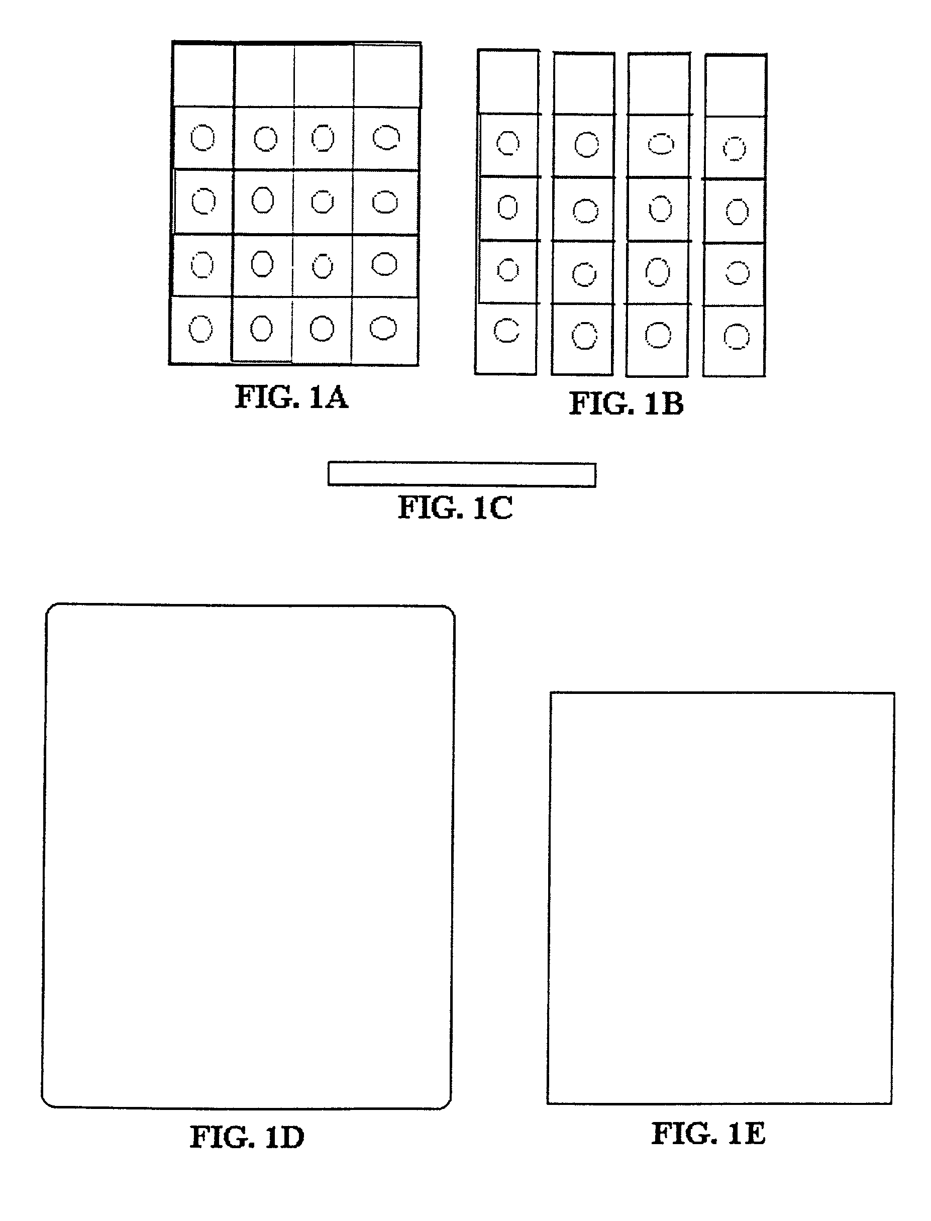
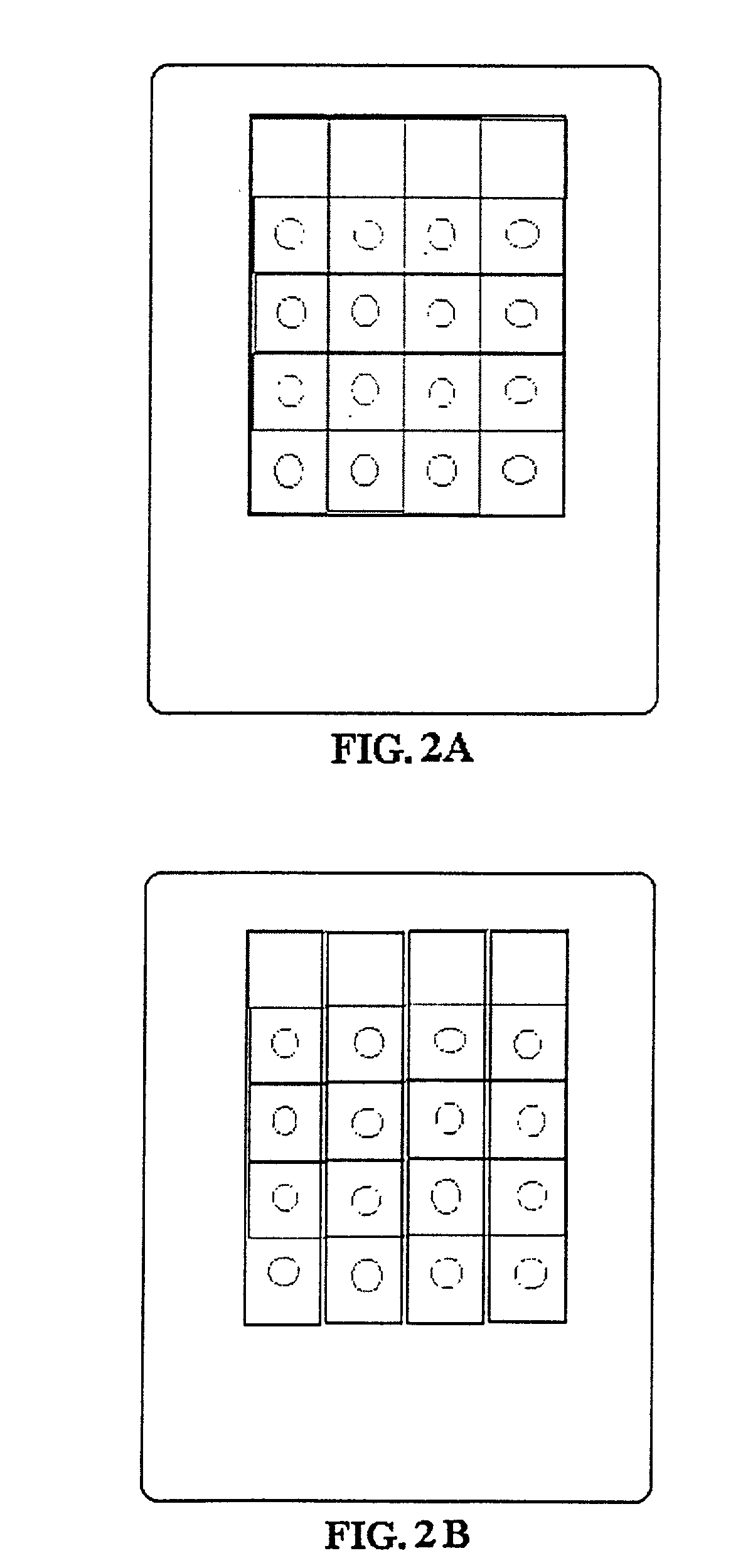
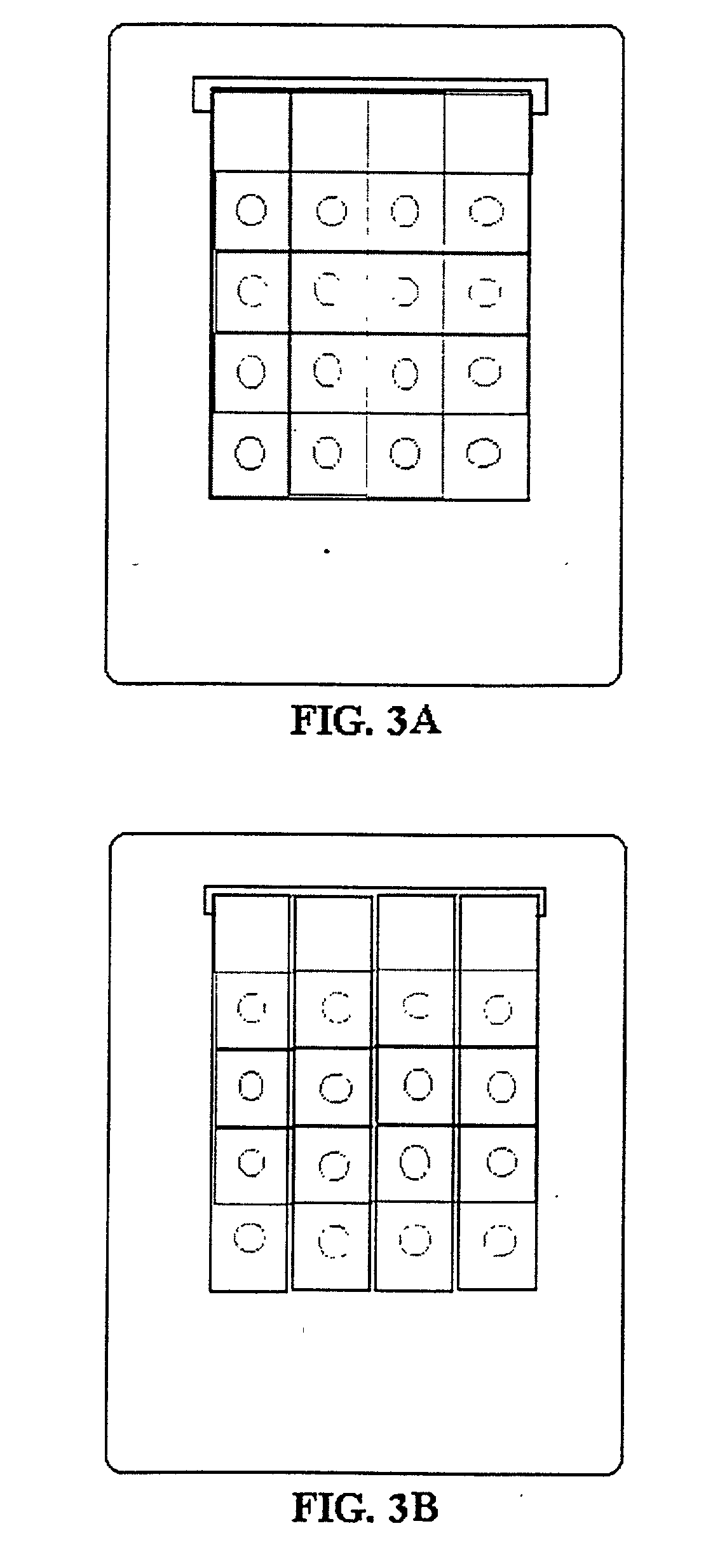
[0098] B. Test Device Preparation. Schleicher & Schuell nitrocellulose membrane having pore size of 0.45 micron is cut to a rectangular piece (14 mm. times. 70 mm) and marked with pencil 14.times.14 mm (FIG. 1A). The membrane can also be cut in strips (14.times.70 mm) and marked with pencil 14.times.14 mm (FIG. 1B). The membrane is then s...

example 2

[0107] The same procedures as Example 1(A), 1(B), 1(C), 1(D), 1(E), 1(F) is followed except that in 1(F) standard 50, 100 and 200 pg / 25 .mu.l were used. Standard or samples is mixed in equal proportion with AFB.sub.1-HRP conjugate (1:500) and 25 .mu.l was added over AFB.sub.1 antibody spotted area. The results obtained in this One-step method were almost similar to that of Two-step.

example 3

[0108] The same procedure as Example 2 is followed except that standard or samples is mixed in equal proportions with 1000-fold AFB.sub.1-HRP conjugate and 25 .mu.l of each mixture were added twice over AFB.sub.1 antibody spotted area. The results obtained in this method were almost similar to those obtained in Examples 1 and 2.


PUM

PropertyMeasurementUnit
Thicknessaaaaaaaaaa
Diameteraaaaaaaaaa
Diameteraaaaaaaaaa

Abstract

A simple easy to manufacture analytical device capable of performing membrane based immunoassays on batch of samples within 3 to 10 minutes wherein the method permits focused application of samples, costly labeled immunoassay and signal amplification reagents, said device includes an antibody-immobilized micro porous membrane, breadth corner layer of which is directly attached to a semi-rigid liquid-impervious body with water insoluble adhesive; absorbent body is provided separately and is not attached to analytical device during manufacture, absorbent body is wetted and is placed proximal to the lower surface of the membrane thereby forming networks of capillary channels with the absorbent body; flow of samples or reagents is always kept downwards and focused without application of any force to the absorbent body and the use of disposable adsorbent body permits stepwise addition of signal amplification reagents for ultra sensitive detection of diagnostically important molecules by visual examination of the membrane surface.

Description

[0001] The present invention relates to analytical device and methods for use in determining analyte in batch of sample suspected of containing such analyte with ultra high sensitivity. More particularly, the invention relates to device and simple methods for application of signal amplification reagents after immunoassay in a laboratory or a field setting for attaining high sensitivity.BACKGROUND AND PRIOR ART INVENTION[0002] The diagnostically important molecules may be small molecules or macromolecules. The small molecules like mycotoxins such as aflatoxin B.sub.1, Ochratoxin A T-2 toxin are present in food and feed such as cereal grains, ground nuts, rice, peanuts, fodder as contaminants due to fungal infection. Human and animals are exposed to mycotoxins through ingestion of toxin-contaminated food and feed, inhalation or skin contact. The determination of mycotoxins in agricultural commodities is important because their consumption by man and animals causes mycotoxicosis. They ...

Claims


Application Information

Patent Timeline
IPC IPC(8): G01N33/543
CPCG01N33/54386Y10S436/809Y10S436/81Y10S436/823
Inventor DHAR, TARUN K.PAL, ARINDAM
Owner COUNCIL OF SCI & IND RES
Learn More