
Methods and apparatus for nonlinear mobility electrophoresis separation

a nonlinear mobility, electrophoresis technology, applied in the direction of liquid/fluent solid measurement, fluid pressure measurement, peptide, etc., can solve the problems of limited separation capability of types of molecules, molecular size and molecular properties, method disclosed,

Inactive Publication Date: 2001-09-27
PROTEOLOGICS INC (US)
View PDF0 Cites 61 Cited by
  • Summary
  • Abstract
  • Description
  • Claims
  • Application Information

AI Technical Summary

Problems solved by technology

Although most commonly used, these electrophoresis techniques have their limitations in separation capabilities regarding types of molecules, molecular sizes and molecular properties.
The method disclosed, however, is limited to charged biomolecules and relatively complicated to run.
Gas chromatography is not suitable to separate and purify biomolecules.
There report also that gradients are predicted to reduce the relative mobilities of the DNA fragments, which is a serious drawback of this technique.
This leads to steep temperature gradients near the end portion of the separation medium which can disturb the separation process.
This leads to steep temperature gradients near the end portion of the separation medium, which can disturb the separation process.
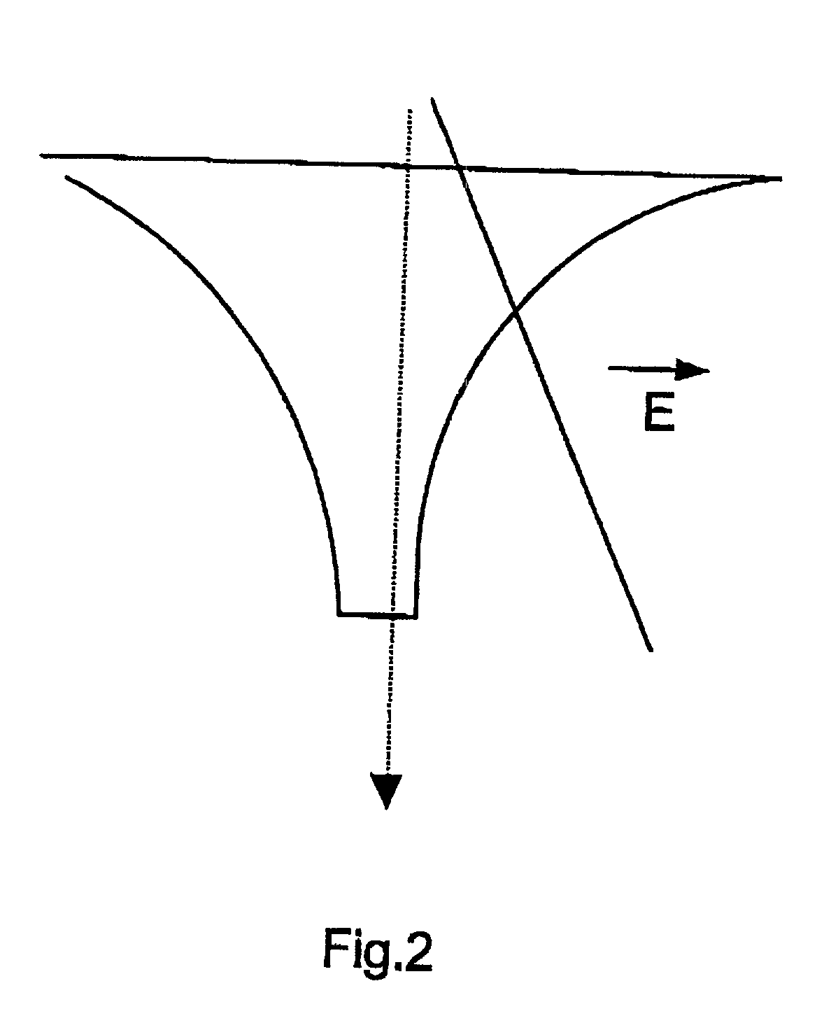
The medium may be limited by limitation means having a shape of two converging hyperbolas.
Special types of electrophoresis cells with two working electrodes and two reference electrodes have been constructed for the generation of non-uniform electric fields, which result in nonlinear mobility focusing.
In variations in which a higher voltage is applied in one direction than in the other, more complex electrical circuitry is required.
Existing methods of separation of large fragments of DNA (>100 kb) based on FIGE and CHEF are slow and tedious.
The existence of non constant fields and of variable media conductivity results in uneven heating of the gel that require additional ways to remove the heat.
Most preparations, however, do contain some anions such as pyruvate and sulfate, which may cause some electro-osmosis.
But Southern blots usually take a long time, frequently 10-20 hours to prepare.
Since the electrophoretic mobility of the H.sup.- is very large the expansion is very rapid and the leading edge very steep.
Reversing of the electric potential will result in a fast decay of the charge wave.
However most problems associated with existing systems are related to the stability of the electric field and thus the stability of proteins running in this field for rectangular shaped vessels.

Method used


Image

  • Methods and apparatus for nonlinear mobility electrophoresis separation
  • Methods and apparatus for nonlinear mobility electrophoresis separation
  • Methods and apparatus for nonlinear mobility electrophoresis separation

Examples


example 2

[0312] Fraction compression in a hyperbolic wedge at constant Potential

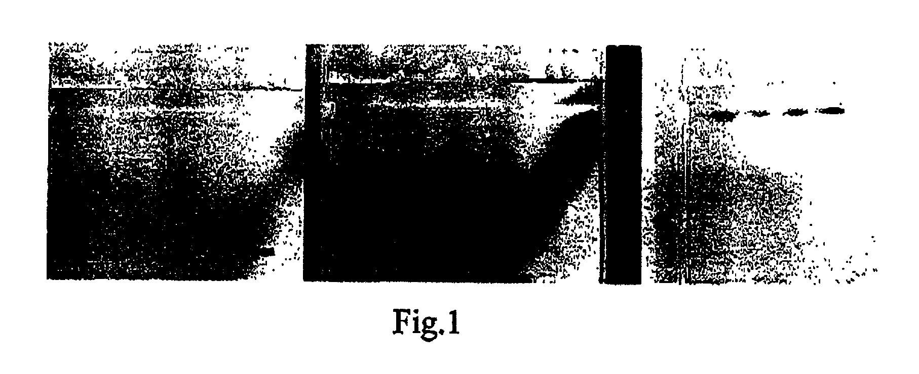
[0313] Two hyperbolic inserts from polymethyl acrilate are placed in the cell with the following characteristics: the length is 13 cm, the width is 10 cm. The insert dimensions vary according to a hyperbolic function with parameters such that the hyperbola narrows from 0 at one end to 4.4 cm at the other end. Agarose gel (0.6%) is poured into the volume between the inserts, creating a gel shaped as a hyperbolic wedge ("hyperwedge") with the length of 8.4 cm and the width of from 10 cm in the widest part to 1.2 cm in its narrow part. The wedge is covered by buffer solution of TAE (height 0.2 cm). The marker DNA lambda-phage is introduced into the start pockets, located at distance of 1.5 cm from the narrow edge. When a constant potential is applied a linearly decreasing field from the narrow to wide end of the hyperbolic wedge provides for focusing of the DNA fractions (see FIG. 25).

example 3

[0314] Nonlinear focusing in the hyperbolic wedge with time varying potential (MEANDER)

[0315] The hyperwedge, which is described in the Example 2, is used for nonlinear DNA focusing. In this Case a variable in time periodic signal of the bipolar pulse wave type (meander) is applied to the electrodes. The average over a period of the electric field is relatively small (less than 2V / cm), in comparison with a field amplitude during the period of the meander action (less than 20V / cm). The sample DNA is entered into the start pockets which are placed in the "narrow" end of the agarose gel (0.6%). Due to nonlinear focusing the more heavy fractions move to the direction of the wide gel end, while the light ones remain behind. With different parameters for the meander voltage the fractions can be made to move to the opposite direction from the start. This behavior differs sharply from all other, traditional methods of electrophoretic DNA separation where everything is determined by the rati...

example 4

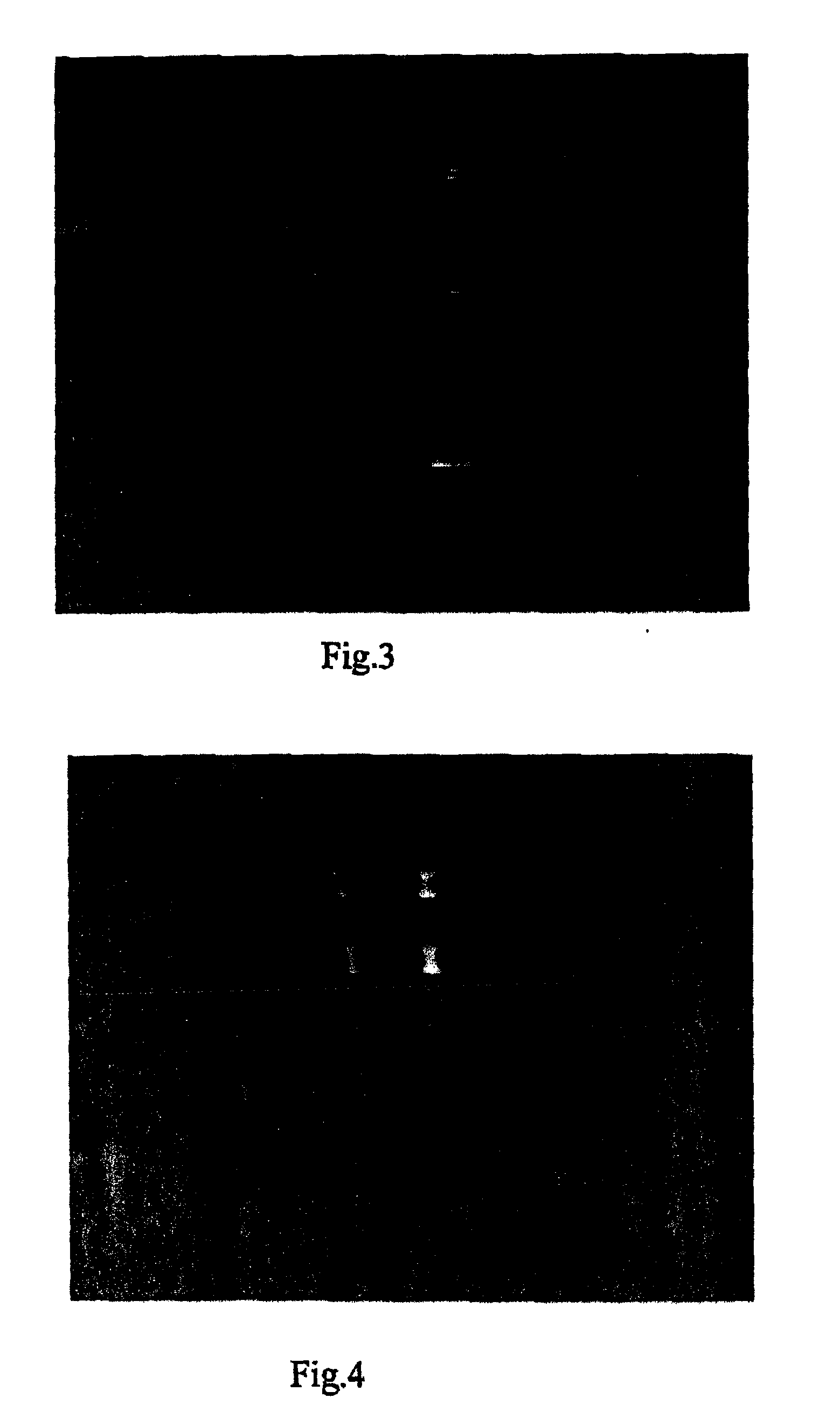
[0316] Consider an electrophoretical cell, partitioned across by non-conductive dielectric walls. There is a hole with the diameter of 50 .mu.m in the wall. The wall divides the cell in to two cameras: right and left one. There is an electrode in each camera, to which a variable in time voltage (AC) is applied with zero average mean voltage. Both cameras near the holes filled with the electrolyte (buffer). A sample, containing a mixture of RBC and hybridoma, is placed into the camera. During the application of a periodic voltage with zero average on the electrodes of the cell, large gradients of the squared electrical field are observed near the hole. Therefore, a dipole power acts on the RBC, forcing them to drift to the area of the strong field. This phenomenon is demonstrated in FIG. 40.

[0317] RBC concentrate near the "hole" when a variable signal is applied. Symmetrical and asymmetrical (FIG. 12) signals were used.

[0318] Separately the nonlinear mobility of RBC was determined by...


PUM

PropertyMeasurementUnit
Timeaaaaaaaaaa
Electric dipole momentaaaaaaaaaa
Timeaaaaaaaaaa

Abstract

A method is disclosed for moving, isolating and / or identifying particles in a sample by placing said sample in a spatially varying electrical field wherein the spatially varying electrical field is following a mathematical nonmonotonous function, selected from the group consisting of linear, hyperbolic, parabolic, parabolic functions or y~xp / q and combinations thereof wherein p q means an integer. Also various devices are disclosed for performing the method.

Description

[0001] The present invention provides a method and apparatus for separation and focusing of cells, cell fractions, chromosomes and molecules, such as nucleic acids and proteins using the effect of nonlinear mobility and / or of non-uniform electric fields.BACKGROUND OF INVENTION[0002] Electrophoresis (EP), dielectrophoresis and electrochromatography are widely used techniques for the analysis, purification, manipulation and separation of mixtures of macromolecules and in particular the study of proteins, nucleic acids and cells. Most applications of electrophoresis are based on the transport of molecules in a supporting gel medium, under the influence of a static electric potential and constant electric field. Many variations of constant field electrophoresis have been employed based on tailoring the electric conductivity and field values by selecting the conductivity properties and pore sizes of the gels, the pH values and the electric potential magnitude. The most important in this ...

Claims


Application Information

Patent Timeline
IPC IPC(8): G01N27/447G01N30/00
CPCG01N27/44773G01N27/44795G01N2030/0035
Inventor FRUMIN, LEONIDPELTEK, SERGEY E.ZILBERSTEIN, GLEB V.BUKSHPAN, SHMUELHALAVEE, URIEL
Owner PROTEOLOGICS INC (US)
Learn More