
Fast bacteria detection and antibiotic susceptibility test by precision tracking of bacterial cells

a technology of bacterial cell and detection method, which is applied in the field of fast bacteria detection, identification (id) and antibiotic susceptibility testing (ast), can solve the problems of one to examine, large optical microscope, large sample preparation time, etc., and achieves the effect of maximal phenotypic feature extraction and minimal sample preparation

Active Publication Date: 2022-08-16
ARIZONA STATE UNIVERSITY
View PDF5 Cites 0 Cited by
  • Summary
  • Abstract
  • Description
  • Claims
  • Application Information

AI Technical Summary

Benefits of technology

This patent describes a new way to detect bacteria using advanced technology. This can be useful in diagnosing infections in patients and determining which antibiotics will work best. The system can also detect bacteria in water, food, and air. Overall, this technology provides a faster and more accurate way to detect and identify bacteria.

Problems solved by technology

The dipstick method is simple and relatively low cost, but it is inaccurate.
However, optical microscopes are bulky, expensive and difficult to use.
This limits one to examine only a small sample volume at a time, which makes it difficult to find bacterial cells in a low concentration sample.
To image bacterial cells in the patient sample, sample enrichment is required, which adds additional difficulty and burden to use the technology.
In addition, traditional optical imaging method requires immobilizing bacterial cells on a surface or in a gel matrix to obtain clear images, which adds extra steps in the sample preparation, and even worse, affects phenotypic features, such as growth and motion, of the bacterial cells.
For these reasons, optical microscopy technology is rarely used by the healthcare provider at the point of care.
While these genotypic approaches are useful, they require a prior knowledge of resistance genes or specific genetic mutations, and are not applicable to phenotypic antibiotic resistance evolved from multiple mechanisms.
These techniques typically require immobilization of bacteria on a surface25, in a gel, or in microfluidic channels for imaging or detection, which raises practical difficulties in testing clinical samples, and affects the growth of the bacterial cells, and thus extraction of growth- and motion-related phenotypical features.
In addition, most of these methods do not work on clinically-relevant concentrations, procedures such as culturing and enriching of the clinical samples, followed by bacterial isolation and purification are still required.

Method used


Image

  • Fast bacteria detection and antibiotic susceptibility test by precision tracking of bacterial cells
  • Fast bacteria detection and antibiotic susceptibility test by precision tracking of bacterial cells
  • Fast bacteria detection and antibiotic susceptibility test by precision tracking of bacterial cells

Examples


example embodiments

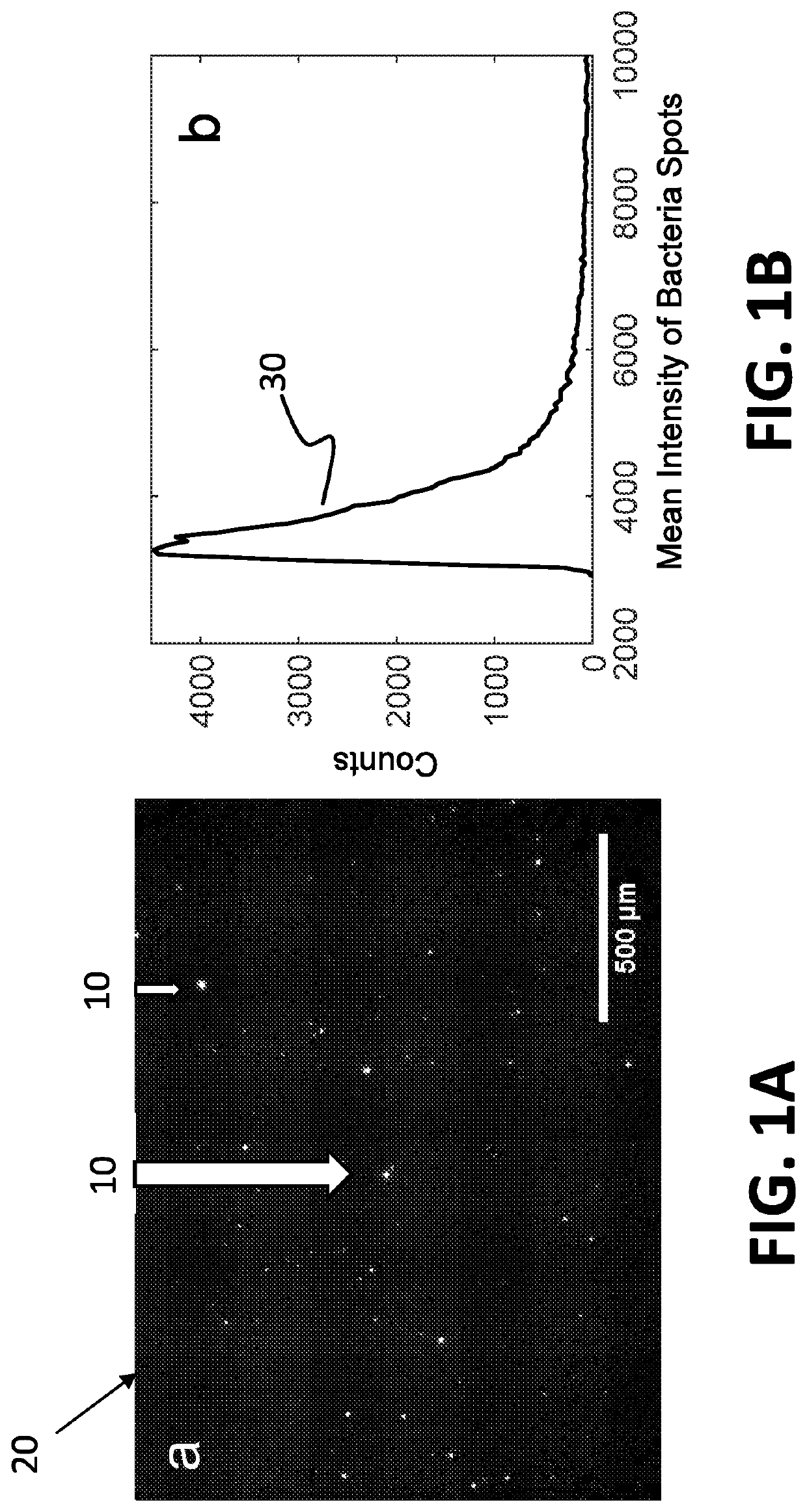
[0062]The system disclosed herein overcomes the drawbacks of the traditional optical microscopy technology with a low spatial resolution imaging system. The imaging system features a large field of view so that it can image a large sample volume and determine the presence and absence of bacterial cells in the patient sample without sample enrichment. For an image volume of 1 μL, the number of bacterial cells is ˜100 for a concentration of ˜105 CFU / mL. The increased imaging volume lowers the spatial resolution of the imaging technology, making it hard to differentiate bacterial cells from other substances in the sample using the traditional imaging analysis algorithm. The present invention describes an apparatus for large sample volume imaging and further discloses algorithms to extract key phenotypic features of bacteria for identification and antibiotic susceptibility test.

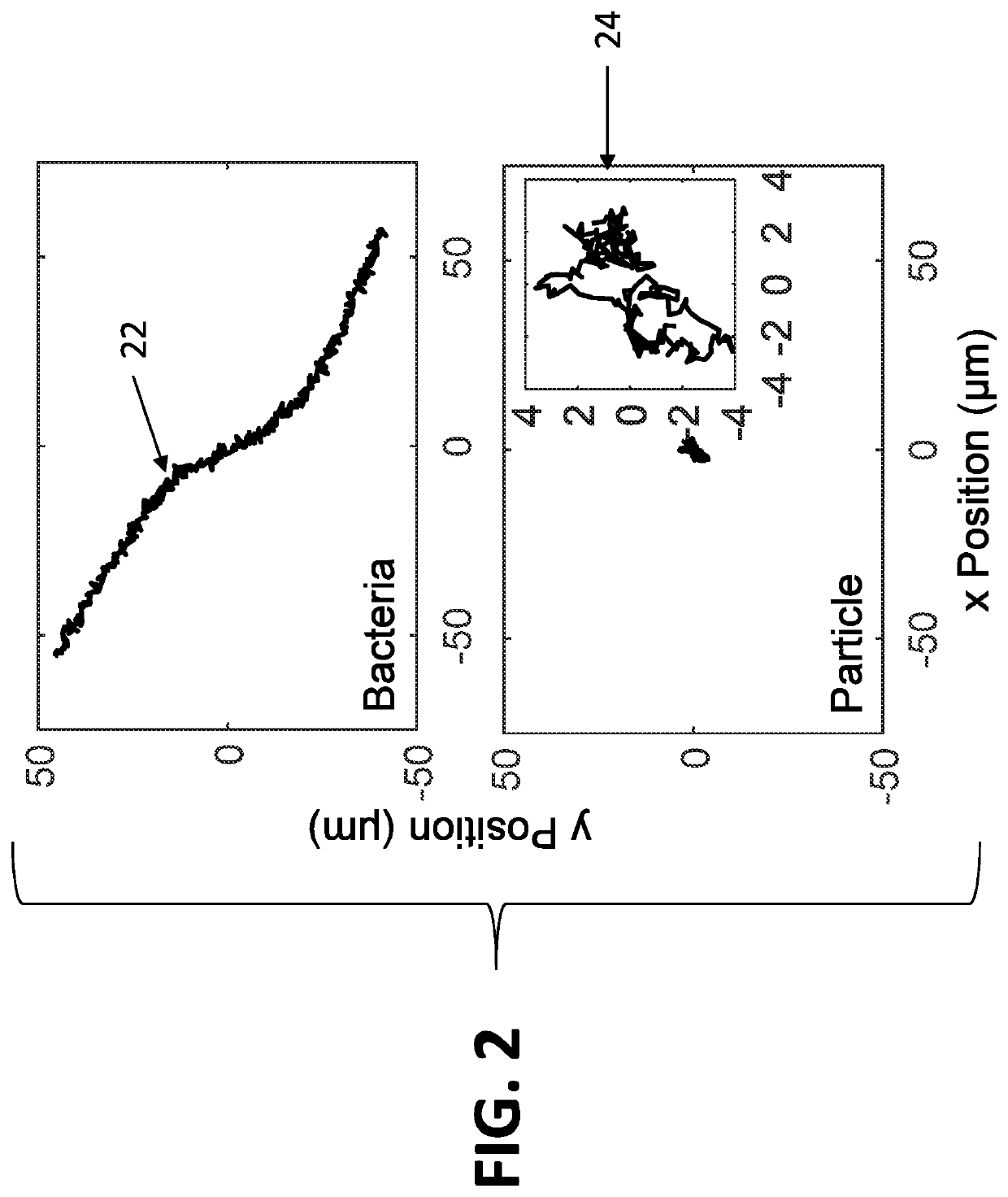
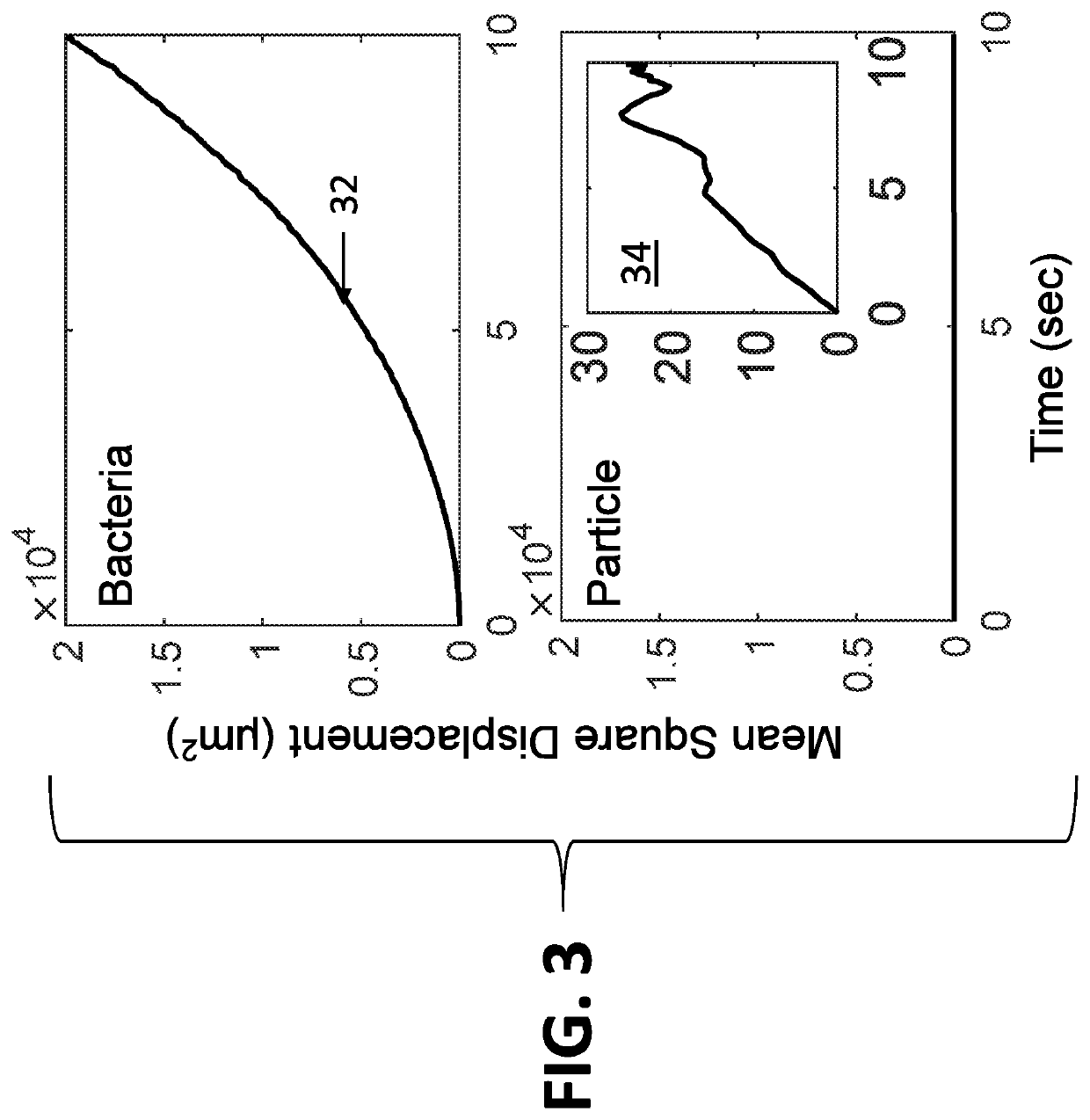
[0063]The phenotypic features include: 1) image intensity of each bacterial cell, 2) position of the cell, and...


PUM

PropertyMeasurementUnit
volumeaaaaaaaaaa
mean square displacementaaaaaaaaaa
volumeaaaaaaaaaa

Abstract

A system for identification of bacterial cells in free solution in a sample. A sample handler is adapted to position the sample. A light source illuminates a large volume of the sample. An imager is located to receive light scattered from the sample. A computer it is coupled to receive data transmitted from the imager. A controller is coupled to send control signals to the sample handler and the computer. The imager processes the scattered light to form images of the bacteria and transmits bacteria image information to the computer, wherein the bacteria image information includes intensity values and position data for the bacteria images from which the computer determines the presence of bacteria.

Description

TECHNICAL FIELD[0001]The present invention relates to fast bacteria detection, identification (ID) and antibiotic susceptibility testing (AST), and, more particularly, to a method and apparatus for fast detection of bacterial cells in free solution using precision tracking of phenotypic features of bacterial cells.BACKGROUND[0002]Timely diagnosis of bacterial infection, and determination of appropriate antibiotic drugs save lives and reduce pain. Currently, healthcare providers often use a dipstick method to identify bacterial infection. The dipstick method is simple and relatively low cost, but it is inaccurate. Optical microscopy obtains a high-resolution image of the patient sample, which is used to identify bacterial cells, and possibly perform antibiotic susceptibility test. However, optical microscopes are bulky, expensive and difficult to use. More importantly, traditional optical microscopy obtains high spatial resolution images of the patient's sample at the expense of prov...

Claims


Application Information

Patent Timeline
Patent Type & Authority Patents(United States)
IPC IPC(8): G06T7/00C12Q1/18G06K9/00H04N5/00G06T7/246G06T7/73G06K9/62H04N5/225H04N5/247G06V10/60G06V10/145G06V10/141H04N23/90
CPCG06T7/0014C12Q1/18G06K9/6201G06T7/246G06T7/73G06V10/145G06V10/60H04N5/2256H04N5/247G06T2207/30024G06V20/693G06V20/695G06V20/698G06V10/141G06F18/22H04N23/56H04N23/90
Inventor TAO, NONGJIANYANG, YUNZE
Owner ARIZONA STATE UNIVERSITY
Learn More