
Composite dura substitute implant

a technology of composite dura and implant, which is applied in the field of composite dura substitute implants, can solve the problems of affecting the growth of bone, affecting the function of the implant, and causing severe consequences, and achieves the effect of facilitating the growth of bone and facilitating the formation of dura

Active Publication Date: 2022-01-11
BEIJING BONSCI TECH CO LTD
View PDF8 Cites 0 Cited by
  • Summary
  • Abstract
  • Description
  • Claims
  • Application Information

AI Technical Summary

Benefits of technology

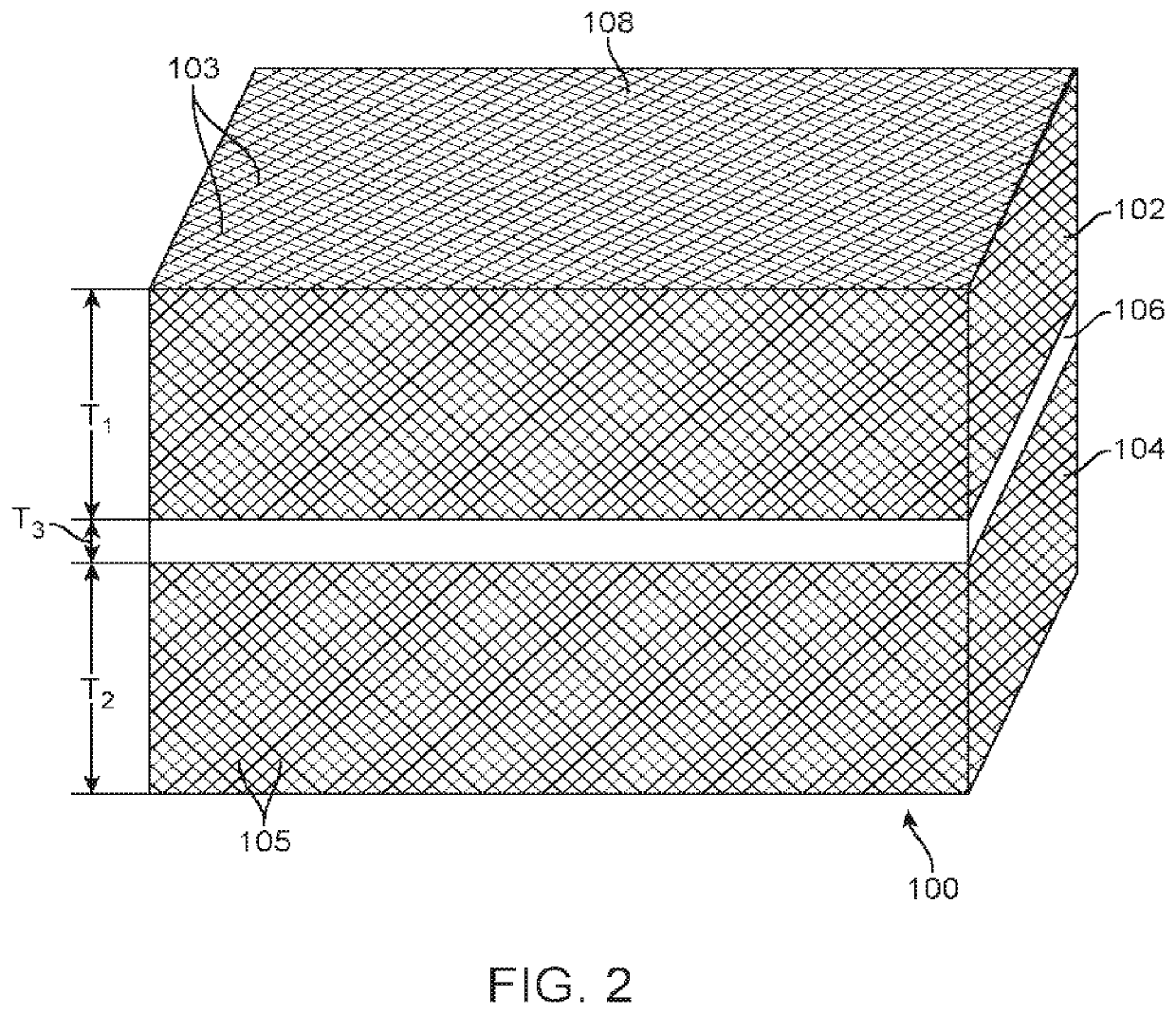
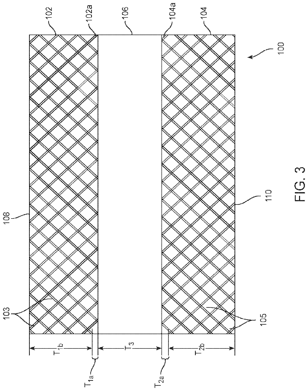
[0009]Embodiment one of the present invention include an implantable dura substitute implant comprising of: a first porous layer comprising a synthetic non-collagen polymer and an osteoconductive material; a second porous layer comprising the synthetic non-collagen polymer and a collagen component; and a middle layer between the first porous layer and the second porous layer, wherein the middle layer is nonporous or has a lower porosity than the first porous layer and the second porous layer, wherein the first porous layer is for contacting bone tissue and the second porous layer is for contacting dura mater upon implantation of the implant at the site of a dural defect in a patient, wherein upon implantation of the osteoconductive material facilitates growth of bone in the first porous layer from the bone tissue and the collagen facilitates dura repair and regeneration in the second porous layer, and wherein the middle layer reduces or prevents leakage of cerebrospinal fluid (CSF) from the second porous layer and dura mater.
[0011]Embodiment two of the present invention includes a method of making a dura substitute implant comprising the steps of: bonding a first porous polymeric layer to a third polymeric layer at a surface of the third polymeric layer; bonding a second porous polymeric layer to the third polymeric layer at an opposing surface of the third polymeric layer, wherein the third polymeric layer is nonporous or has a porosity sufficiently low that upon implantation of the implant the third polymeric layer prevents leakage of cerebrospinal fluid; and impregnating the first porous polymeric layer with a osteoconductive material; and impregnating the second porous polymeric layer with collagen.
[0013]Embodiment three of the present invention includes a method of treating a dural defect in a patient having damaged dura mater comprising: implanting a dura substitute implant on a site of the dural defect of the brain of the patient, wherein the dural defect is in contact with a bottom porous layer of the implant comprising a synthetic non-collagen polymer and collagen and the collagen facilitates dura repair and regeneration tissue within the bottom porous layer, wherein bone tissue is in contact with a top porous layer of the implant comprising the synthetic non-collagen polymer and an osteoconductive calcium phosphate material which causes bone growth in the top porous layer and integrates the top porous layer with cranial tissue, and wherein leakage of cerebrospinal fluid from the site is prevented by an intervening layer between the top and bottom porous layers comprising the synthetic non-collagen polymer. One may want the bony ingrowth or an osteoconductive structure to be a thickness of 1 mm or thicker.

Problems solved by technology

When the integrity of a person's meningeal membranes is intentionally or accidentally compromised, serious consequences may ensue, unless the membranes can be repaired.
Defects of the dura mater or dura defect is a common clinical issue resulting from trauma, inflammation, tumor resection and congenital malformations.
Complications such as cerebrospinal fluid leakage, infections, meningitis, and epilepsy can be generated due to inadequate closures of the dura after neurosurgery.
Defects of the dura mater can produce a variety of undesirable consequences such as brain herniation, adhesion formation between the neural tissue and the overlying structures, pseudomeningocele, cortical scarring, cerebrospinal fluid fistulas and wound infection with potential propagation to the brain parenchyma.
There are a number of issues with current dura substitute implants.
Both autografts and allografts have limited supply.
The risk of transmission of the CJD agent has led to the ban of cadaveric dura mater products.
Non-degradable synthetic materials are bioinert and do not integrate well with tissues.
Degradable synthetic materials typically generate acidic by-products which may induce inflammation reactions.
In addition, degradable synthetic materials may either degrade or lose mechanical strength too fast or too slowly.
Both non-degradable and degradable synthetic materials have been rejected clinically because of local tissue reactions, excessive scar formation, meningitic symptoms, or hemorrhage risk.
However, such xenogenic materials are nonresorbable, and may result in a risk of complications associated with infection and immunorejection.
Degradable collagen matrices based on bovine tendons have raised concerns of rate of degradation, low mechanical strength and usage with sutures.

Method used


Image

  • Composite dura substitute implant
  • Composite dura substitute implant
  • Composite dura substitute implant

Examples


example 1

[0070]An example of a composite dura substitute implant is composed of three layers of PEKK material:[0071]The top layer is PEKK fabric is coated with bone-like apatite, has a porosity of 50%, and thickness of 2 mm.[0072]The middle layer of PEKK fabric has a porosity less than 0.1% and thickness of 0.5 mm.[0073]The bottom layer is PEKK fabric is coated with type I collagen, has a porosity of 50%, and thickness of 1 mm.

example 2

[0074]An example of a composite dura substitute implant is composed of three layers of UHMWPE material:[0075]The top layer is Dyneema® fabric offered by DSM company, is coated with bone-like apatite, has a porosity of 60%, and thickness of 2 mm.[0076]The middle layer is a UHMWPE sheet with a porosity less than 0.1% and thickness of 0.5 mm.[0077]The bottom layer is Dyneema® fabric coated with type I collagen, has a porosity of 60%, and thickness of 1.5 mm.

example 3

[0078]An example of a composite dura substitute implant is composed of three layers of PEEK material:[0079]The top layer is PEEK fabric coated with bone-like apatite, has a porosity of 50% and thickness of 1.5 mm.[0080]The middle layer is a PEEK sheet with a porosity less than 0.1% and thickness of 0.5 mm.[0081]The bottom layer is PEEK fabric coated with type I collagen, has a porosity of 50%, and thickness of 1 mm.


PUM

PropertyMeasurementUnit
porosityaaaaaaaaaa
porosityaaaaaaaaaa
porosityaaaaaaaaaa

Abstract

A composite dura substitute implant for implantation at a dura defect site having a porous layer that provides an osteoconductive scaffold for bony ingrowth, a porous layer that provides a scaffold for regeneration of collagen at a dura surface, and an intervening layer for preventing cerebrospinal leakage is disclosed. The composite dura substitute implant facilitates regeneration of dura mater and promotes osteointegration with bony tissue. Methods of manufacturing such an implant and methods of treatment using such composite dura substitute implants are further disclosed.

Description

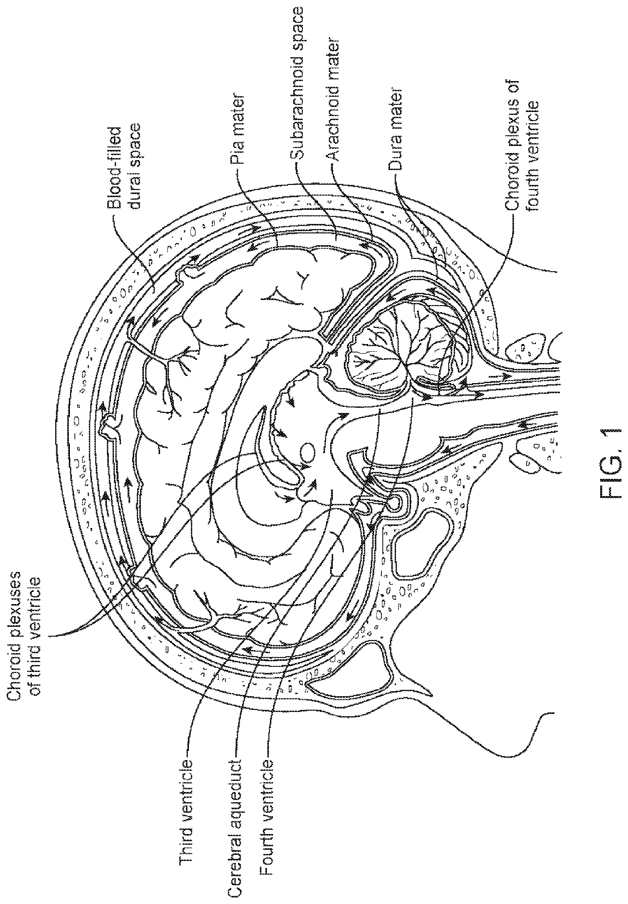
[0001]This application is a divisional of application Ser. No. 16 / 015,166 filed on Jun. 21, 2018, which is a continuation of application Ser. No. 15 / 405,066 filed on Jan. 12, 2017 and claims priority to Chinese Patent Application No. 201610884151.7 filed on Oct. 10, 2016 each of which is incorporated by reference herein.BACKGROUND OF THE INVENTIONField of the Invention[0002]This application is directed to composite dura substitute implant to repair and regenerate damaged dura tissue. More particularly, this application is directed to composite dura substitute implant formed of joined layers of porous bodies, methods of forming the same, and methods of treatment.Description of the State of the Art[0003]The human brain and spinal cord are covered with meningeal membranes whose integrity is critical to the operation of the central nervous system. FIG. 1 depicts an illustration of the human brain. In particular, the meningeal membrane comprises three overlapping layers of tissue, which ...

Claims


Application Information

Patent Timeline
Patent Type & Authority Patents(United States)
IPC IPC(8): A61L27/56A61L27/36A61L27/48A61L27/46
CPCA61L27/56A61L27/3675A61L27/46A61L27/48
Inventor NIE, HONGTAOZHANG, KAI
Owner BEIJING BONSCI TECH CO LTD
Learn More