Endoscopic system and methods having real-time medical imaging

Active Publication Date: 2021-12-07
SATISFAI HEALTH INC
View PDF30 Cites 3 Cited by
  • Summary
  • Abstract
  • Description
  • Claims
  • Application Information

AI Technical Summary

Benefits of technology

The invention is a system that uses artificial intelligence to detect polyps in a 2D image of a colon and create a 3D model of the colon. The system also highlights areas that may be overlooked during colonoscopy and helps the endoscopist go back and image those areas. The system also includes a storage and indexing module for recording and storing the 3D model and 2D image for comparison during a subsequent examination. The system includes multiple display windows to show real-time images and an overlay of information generated by the artificial intelligence module. The system also includes a navigation module to help with orientation and navigation during the procedure. The 2D flat image of the anatomy provided in near real-time on the second display window also may be analyzed by the artificial intelligence model to enhance the detection of polyps.

Problems solved by technology

One factor is the difficulty of identifying a polyp or adenoma, even though it may be in the visual field of the colonoscopy image.
Another factor that contributes to the lower than ideal ADR is the difficulty of ensuring that the entire internal surface of the colon has been imaged.
It is difficult for a colonoscopist to remember what has been imaged, and “integrate” those images mentally to conclude that the entire internal surface has been examined.
Thus, it is challenging for the endoscopist to ensure that the entire internal surface of the colon has been visualized.
Failure to visualize the entire internal surface poses a risk of missing potentially harmful polyps or cancers.
One of the challenges of endoscopy, particularly colonoscopy, is that the endoscopist views the structure of interest in real time with only a forward-looking view from the tip of the endoscope.
Moreover, the colon is a constantly moving organ that lacks rigid boundaries, so insertion of a colonoscope can cause changes in the shape and configuration of the colon.
Further, the folds in the wall of the colon are constantly moving, thus making it very difficult for an endoscopist to ensure that every fold, nook and cranny is visualized during an examination.
Yet another challenge in endoscopy is that the tip of the endoscope is controlled by a series of knobs and switches located on the hand piece manipulated by the endoscopist.
Accordingly, it is quite difficult for many endoscopists, especially those for whom endoscopy is not part of their daily practice, to translate the image on the screen into necessary hand movements to re-direct the tip of the endoscope.
Hence, control reference between the tip of the endoscope and the controls on the hand piece may be difficult to maintain.
A limitation of the foregoing approach is that the image presented to the clinician involves interpolation and extrapolation of data from a true imaging modality, resulting in loss of resolution, features and formal correctness.
Moreover, through-body imaging modalities, such as X-ray, CT scanning and MRI have fundamental resolution limits such that small features may be missed.
In general, these historical approaches employ “post processing” of images after an examination is completed, and cannot provide real time information while the clinician is performing the examination.
In addition, such previously known 3D reconstructions lack registration with the actual anatomy, and thus cannot provide precise positional information for a later-performed procedure.
For example, a clinician performing a procedure while viewing a 3D reconstruction generated during a prior examination, typically will find the live anatomy to be distorted, moved, squished and / or stretched compared to the prior 3D reconstruction.
This lack of temporal continuity is a significant limitation of existing methods of 3D reconstruction of anatomy.
For example, fiducial markers created using gamma radiation have been proposed to provide some level of registration, but that technique exposes a patient to an additional dose of gamma radiation, which is itself potentially harmful, as described, for example, in Kleiman N J, Macvittie T J, Aleman B M, Edgar A B, Mabuchi K, Murihead C R, Shore R E, Wallace W H.
Although that approach has some value, it includes all of the foregoing mentioned limitations, including low resolution and difficulty in finding an identified area during a later examination.
If a polyp to be removed is identified on virtual colonoscopy, then at least one additional procedure is required to remove the polyp, and finding the corresponding area often is challenging for all of the reasons mentioned above.
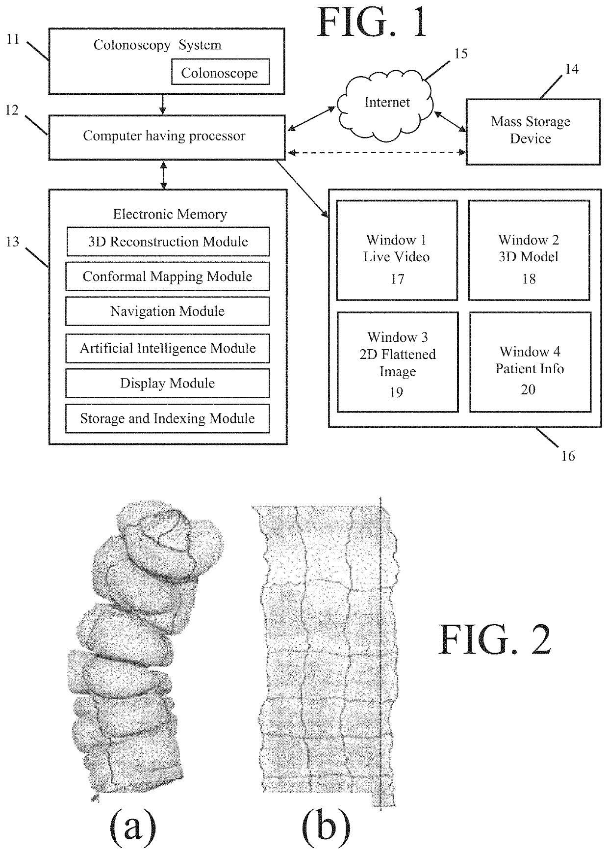
Those patents do not describe how to solve the problems highlighted above, nor do they suggest systems or methods for generating an evolving 3D or flat 2D image without reference to a prior set of images.
However, the approach described in that article suffers from the limitation that it is trained on synthetic data from a physical model, not real-world images, and then validated on a porcine colon but with CT registration.
The resulting system algorithms are neither independent nor capable of providing real time information.
Such 3D colon reconstruction techniques cannot be used during a procedure, and so do not address the problems sought to be addressed by this disclosure.
That patent does not teach a method or system for performing the same function on images obtained in real time to provide guidance to an endoscopist during an optical colonoscopy procedure.
Another challenge of colonoscopy involves detection of polyps and other features in real time.
That patent does not address any of the issues of 3D reconstruction, creation of a 2D flat image, or real time registration discussed above.
The authors noted that their system is only able to work in “chunks” because the system fails on large camera motions or deformation.
In addition, no previous system or method provides for an overlay on such images of AI detected features of interest, such as polyps.

Method used


Image

  • Endoscopic system and methods having real-time medical imaging
  • Endoscopic system and methods having real-time medical imaging
  • Endoscopic system and methods having real-time medical imaging

Examples


Embodiment Construction

[0048]The present invention provides systems and methods for improving endoscopy procedures by enhancing the endoscopists' viewing, navigation and interpretation of the interior surface of an organ being examined. In this disclosure, an exemplary system is described for performing colonoscopy, wherein the system provides not only a conventional real time image of the view obtained by the colonoscope, but in addition, a near real time 3D model of the colon and / or a 2D flat image of the interior surface of the colon. In accordance with one aspect of the invention, the real time 3D model and / or 2D image are processed using AI software, which highlights potential features, e.g., polyps or potential adenomas, for closer examination and / or biopsy during the procedure. A navigation module interacts with other system outputs to further assist the endoscopist with navigational indicia, e.g., landmarks and / or directional arrows, that enhance the endoscopists' spatial orientation, and / or may p...


PUM


Abstract

Systems and methods for improving endoscopy procedures are described that provide not only a conventional real time image of the view obtained by an endoscope, but in addition, a near real time 3D model and / or a 2D flattened image of an interior surface of an organ, which model and image may be processed using AI software to highlight potential tissue abnormalities for closer examination and / or biopsy during the procedure. A navigation module interacts with other system outputs to further assist the endoscopist with navigational indicia, e.g., landmarks and / or directional arrows, that enhance the endoscopists' spatial orientation, and / or may provide navigational guidance to the endoscopist to assist manipulation of the endoscope.

Description

FIELD OF THE INVENTION[0001]The present invention relates generally to the field of real-time 3 dimensional (3D) reconstructed and 2D flattened imaging of a body cavity, with particular application to endoscopy such as colonoscopy.BACKGROUND OF THE INVENTION[0002]Endoscopy refers to a medical procedure in which an instrument is used for visual examination of an internal body part. A common example of endoscopy is colonoscopy, during which a flexible tube with imaging apparatus at the distal end is inserted into a person's colon. The purpose of colonoscopy is to search for and identify abnormalities in the internal wall of the colon and, in some cases, remove them. Such abnormalities include polyps and various types of adenomas.[0003]Screening colonoscopy remains the best-proven method to detect and prescribe early treatment for colon cancer. Clinical guidelines typically suggest that a first colonoscopy be performed at age 50. In screening colonoscopy, the colonoscopist performs a r...

Claims


Application Information

Patent Timeline
IPC IPC(8): G06K9/00A61B1/00A61B1/31A61B1/01G06T7/00G06N20/00G06T1/60
CPCA61B1/0005A61B1/01A61B1/31G06N20/00G06T1/60G06T7/0016G06T2207/10068G06T2207/30028G06T7/0012G06T19/00G06T2210/41G06T2219/028G06T17/00G06T2200/24A61B1/000096A61B1/00194
Inventor ZINGARETTI, GABRIELECROSBY, PETERNINH, ANDREWREQUA, JAMESKARNES, WILLIAM E.CIFARELLI, JOHN
Owner SATISFAI HEALTH INC
Why Eureka
Try Eureka