Method for font printing-preparation of tissue slice, and fill-up of tissue chip

A sectioning and histological technology, applied in the field of sectioning with carrier adhesion, can solve the problems of tissue point inconsistency, low pass rate of tissue chip products, falling off, etc.

Active Publication Date: 2005-11-16
SHANG OUTDO BIOTECH CO LTD
View PDF0 Cites 6 Cited by
  • Summary
  • Abstract
  • Description
  • Claims
  • Application Information

AI Technical Summary

Problems solved by technology

The problem of "invalid tissue": Before the tissue chip is made, the tissue wax block must be diagnosed and marked. At this time, only the surface of the tissue is diagnosed and marked, and the structural changes in the deep tissue cannot be detected through the surface, such as a In the tumor tissue core, some are tumor tissue, some are tumor necrosis tissue called "ineffective tissue", fibers, blood vessel tissue, etc., which often cause some tissue points to be inconsistent with the original template design and original diagnosis. This phenomenon is almost It is unavoidable and can appear in all tumor tissues, and chips with too many "invalid tissues" can only be treated as waste chips
The problem of lack of displacement of sample tissue points: There are many procedures for making tissue microarrays, and the length of tissue cores is different, the tissue core and the acceptor wax block are not tightly combined, and the tissue core may shift or even fall off when slicing.
These two problems make the qualification rate of tissue chip products very low

Method used


Image

  • Method for font printing-preparation of tissue slice, and fill-up of tissue chip
  • Method for font printing-preparation of tissue slice, and fill-up of tissue chip
  • Method for font printing-preparation of tissue slice, and fill-up of tissue chip

Examples


example 1

[0023] Example 1 Tissue chip slice preparation

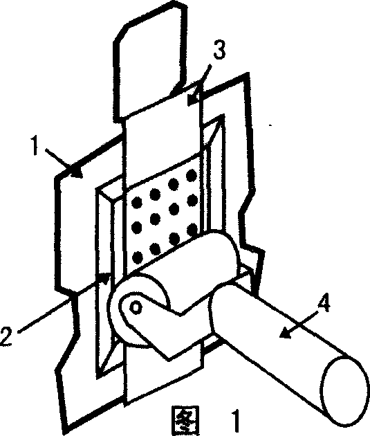
[0024] The first step is shown in Figure 1: the paraffin tissue array block (2) is fixed on the sample holder (1) of the microtome, and the surface of the sample is cut and trimmed with the microtome in the trimming mode to fully expose the target, and a section with organic adhesion is taken. Adhesive adhesive tape (3) (the area is slightly larger than the target), remove the back bottom plate, stick to the surface of the target through the adhesive on the tape, and gently roll the film with the rubber roller (4) to make the two are tightly adhered.

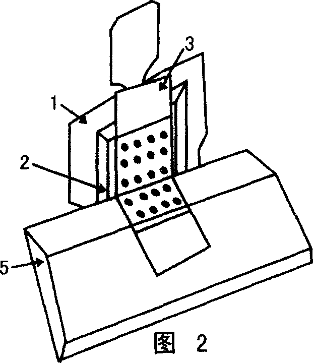
[0025] The second step is shown in Figure 2: use the tissue microtome blade (5) to remove the adhesive tape (3) with the tissue chip attached from the paraffin tissue array block ( 2) The surface is cut off.

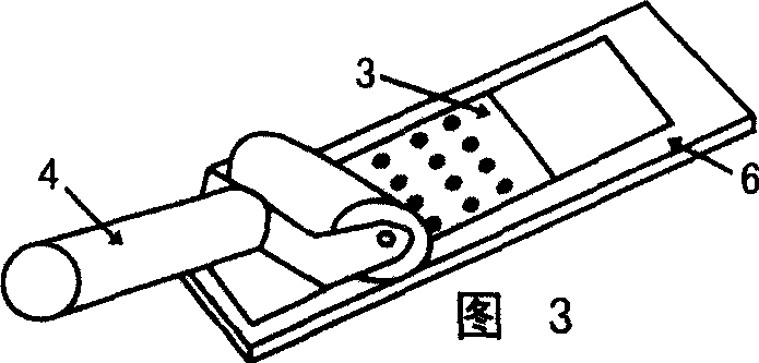
[0026] The third step is shown in Figure 3: stick the adhesive tape (3) with the tissue chip section on the passing glass slide (6), and roll it with a rubber roller (4) t...

example 2

[0028] Example 2 is used to fill in missing tissue samples in tissue microarrays

[0029] This method can also be used to repair missing tissue samples in tissue microarrays, that is, the movable type repair method.

[0030] The first step is as Figure 6 Shown: Tissue cores are extracted from pathological wax blocks in the process of making tissue microarrays. The thickness of pathological wax blocks is different, and the length of tissue cores is also different. Slicing tissue cores with different lengths into tissue arrays may result in the loss of Some sample points are missing, and mistakes may also occur when extracting tissue cores. The sample points are different from the actual array design. We can remove the wrong sample points to form a tissue microarray with missing parts.

[0031] The second step is shown in Figure 7: arrange the tissue cores to fill the sample points in a blank wax block according to the array design, and fix the wax block (11) containing the s...


PUM


Abstract

A movable type printing type method for preparing and repariing the tissue chip includes sticking the chip substrate carrying organic adhesive onto the surface of wax block, slicing, sticking the sliced section onto glass plate, baking, cooling, and putting it in organic solvent to dissolve said adhesive and wax.

Description

1. Technical field [0001] The present invention relates to a histological slicing method, more particularly, relates to a slicing method utilizing carrier adhesion. 2. Background technology [0002] With the development of histology and pathology, people's understanding of the human body or other animals and plants has developed from the anatomical organ level to the tissue and cell level. However, the observation of tissues and cells needs to be carried out through microscope tools. However, since the complete organ is difficult to observe with a microscope, it is necessary to cut the organ into very thin sections for staining and observation. In order to solve this problem, a tissue section method has emerged. The principle of this method is to cut the organ into small pieces of tissue, fix it with formalin, use alcohol to remove the moisture in the tissue, then immerse it in xylene or benzene solution to remove the alcohol, and finally immerse the tissue piece in molten ...

Claims


Application Information

Patent Timeline
IPC IPC(8): B41M1/30
Inventor 郜恒骏张宝峰姚海嵩
Owner SHANG OUTDO BIOTECH CO LTD
Why Eureka
Try Eureka