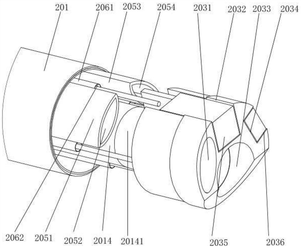
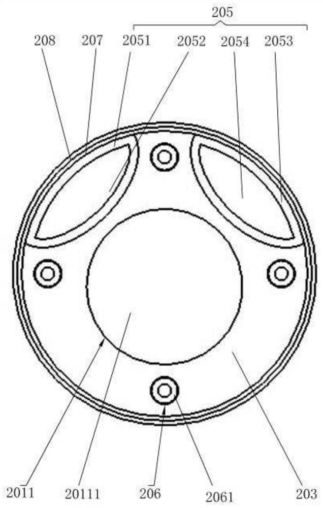
Medical catheter and medical device
A technology of medical catheters and outer tubes, applied in catheters, other medical devices, applications, etc., can solve problems such as head displacement, insufficient lighting, and prolong operation time, reduce sampling times, overcome insufficient lighting, and expand the scope of use Effect
- Summary
- Abstract
- Description
- Claims
- Application Information
AI Technical Summary
Problems solved by technology
Method used
Image


Examples
Embodiment 1
[0150] The working process of the inner control and bending mechanism described in the first embodiment is as follows:
[0151] Rotating the locking handle 145, the locking handle 145 rotates and drives the pressing rod 144 to rotate and moves downward in a small range through the thread. The distal ends of the adjacent locking pieces 147 are closed relative to each other and hold the upper wheel 127 and the lower wheel 119 respectively, so as to realize the self-locking of the medical catheter 2 in any direction. In the non-self-locking state, the distal ends of the adjacent locking pieces 147 can be relatively opened by the spring 148 and contact the self-locking of the upper wheel disc 127 and the lower wheel disc 119 .
[0152] The specific structure of the second embodiment of the inner control bending self-locking mechanism is described below. The inner control bending self-locking mechanism is provided outside the handle body 1 .
[0153] Please refer to Figure 15 an...
Embodiment 2
[0156] The working process of the inner tube-controlled bending locking mechanism described in the second embodiment is as follows:
[0157] Please refer to Figure 15 and Figure 16 , the rotation locking knob 109 drives the rotation limit block 140 to rotate, and the rotation limit block 140 rotates along the surface 1411, so that the fixed limit block 141 is subjected to the pressing force in the axial direction to realize the axial movement. The back of the block 141 is provided with a first locking washer 1421, the first locking washer 1421 is squeezed and presses the lower wheel 108 in the axial direction, and the lower wheel knob 108 is moved by the pressing force, During the moving process, the first locking washer 1421 is compressed and deformed. After the first locking washer 1421 is deformed, the damping is increased and the shaft end of the lower disc 119 is held tightly so that it cannot rotate. At the same time, after the lower roulette knob 108 is squeezed, th...
Embodiment 3
[0172] The specific structure of the inner tube locking mechanism 101 described in the third embodiment is as follows:
[0173] The inner tube locking mechanism 101 includes a second fixing part and a second rotating part hinged with the second fixing part, and the second fixing part and the other end of the second rotating part that are not connected through a tight Fixed device connection.
[0174] Please refer to Figure 22 , Specifically, the second fixed part adopts a fixed end 10112, and the second rotating part adopts a clamping end 10110. The fixed end 10112 and the clamping end 10110 are respectively provided with semicircular shapes for matching the outer diameter of the medical catheter 2. In the clamping port 10114, one end of the fixed end 10112 and one end of the clamping end 10110 are hinged through a hinge 10111, and the other end of the fixing end 10112 and the other end of the clamping end 10110 are respectively provided with threaded holes. The above-menti...
PUM
 Abstract
Description
Claims
Application Information
 - R&D
- Intellectual Property
- Life Sciences
- Materials
- Tech Scout
- Unparalleled Data Quality
- Higher Quality Content
- 60% Fewer Hallucinations
Browse by: Latest US Patents, China's latest patents, Technical Efficacy Thesaurus, Application Domain, Technology Topic, Popular Technical Reports.
© 2025 PatSnap. All rights reserved.Legal|Privacy policy|Modern Slavery Act Transparency Statement|Sitemap|About US| Contact US: help@patsnap.com