
Parathyroid gland identification method based on image fusion technology

A parathyroid gland and image fusion technology, applied in the field of medical assistance, can solve the problems of difficulty in guaranteeing the recognition accuracy and interference of autofluorescence imaging, and achieve the effects of avoiding missed detection, improving accuracy, and improving safety

Pending Publication Date: 2022-03-11
高永楷
View PDF0 Cites 0 Cited by
  • Summary
  • Abstract
  • Description
  • Claims
  • Application Information

AI Technical Summary

Problems solved by technology

However, in actual use, it was found that some lymph nodes and adipose tissue after electrocautery will also produce autofluorescence, causing interference, which makes it difficult to guarantee the accuracy of autofluorescence imaging identification

Method used


Image

  • Parathyroid gland identification method based on image fusion technology

Examples


Embodiment

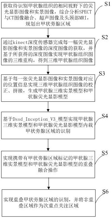
[0031] Such as figure 1 As shown, a method for identifying parathyroid glands based on image fusion technology includes the following steps:

[0032] S1. Obtain the fluorescein imaging image and the real scene image under the same field of view of the thyroid tissue to be identified, comprehensively analyze the fusion of SPECT and CT images, ultrasound images and MRI of the head and neck, and plan the parathyroid gland area; The thyroid tissue image acquired by the imaging surgical navigation system, the real scene image is the thyroid tissue image acquired under the same field of view through a laparoscope;

[0033] S2. Complete the acquisition of the depth images of each fluoroscopy image and real-scene image through the kinect depth sensor, and realize the three-dimensional reconstruction of the thyroid tissue image based on the obtained depth image, and obtain the three-dimensional thyroid tissue image; specifically, through the kinect depth The sensor completes the acqui...


PUM


Abstract

The invention discloses a parathyroid gland identification method based on an image fusion technology, and the method comprises the following steps: obtaining depth images of a fluorescent development image and a live-action image, and achieving the three-dimensional reconstruction of a thyroid tissue image based on the depth images, thereby obtaining a three-dimensional thyroid tissue image; correcting and splicing the three-dimensional thyroid tissue image based on the position information corresponding to the fluorescence development image and the real scene image, and generating a thyroid three-dimensional real scene model and a thyroid fluorescence development model; on the basis of a DssdInceptionV3 model, identification of parathyroid gland areas loaded in the thyroid three-dimensional live-action model and the thyroid fluorescence development model is achieved; overlapping fusion operation of the thyroid three-dimensional live-action model carrying parathyroid gland area marks and the thyroid fluorescence development model is realized; recognition of an overlapped parathyroid gland area is realized, and a non-overlapped area is used as a secondary focus area. According to the invention, the parathyroid gland identification accuracy is greatly improved.

Description

technical field [0001] The invention relates to the field of medical auxiliary technology, in particular to a method for identifying parathyroid glands based on image fusion technology. Background technique [0002] The parathyroid gland is an important endocrine gland in the human body, and its main function is to secrete parathyroid hormone (PTH for short). The living parathyroid glands are generally oblate and oval, with a yellow or brownish-yellow appearance, resembling soybeans. Under normal circumstances, each person has four parathyroid glands, which are symmetrically distributed in the middle and lower parts of the back of the left and right thyroid glands, but some people have large differences in the location and number of parathyroid glands. [0003] The parathyroid glands are small in size and their positions and numbers are not fixed, making it difficult to distinguish them from ectopic thyroid, ectopic thymus, surrounding fat, and lymph node tissue, which grea...

Claims


Application Information

Patent Timeline
IPC IPC(8): G06T7/00G06T7/73G06T5/50G06T17/00G06T19/20
CPCG06T7/0014G06T7/73G06T5/50G06T17/00G06T19/20G06T2207/10121G06T2207/10081G06T2207/10108G06T2207/10132G06T2207/10088G06T2207/20221G06T2210/41G06T2210/61
Inventor 高永楷
Owner 高永楷
Learn More