Medical image processing method and device, terminal and storage medium

A medical image and processing method technology, applied in the field of medical assistance, can solve the problems of low specificity and accuracy, complex judgment process of esophageal marker infiltration depth, and difficult to guarantee the accuracy of esophageal marker infiltration depth estimation, etc., to achieve specificity The effect of improving the accuracy and improving the accuracy

Active Publication Date: 2022-02-22
WUHAN UNIV
View PDF4 Cites 0 Cited by
  • Summary
  • Abstract
  • Description
  • Claims
  • Application Information

AI Technical Summary

Problems solved by technology

Based on the above features related to the depth of esophageal markers, the process of judging the depth of esophageal marker infiltration is complicated. Due to the heterogeneity of clinical experience and subjective judgment of endoscopists, the accuracy of preoperative infiltration depth estimation of esophageal markers Difficult to guarantee
[0004] However, the current determination of the infiltration depth of esophageal markers has low specificity and accuracy of the results. Therefore, how to improve the specificity and accuracy of medical image processing is a technical problem that needs to be solved urgently in the field of medical assistance technology

Method used


Image

  • Medical image processing method and device, terminal and storage medium
  • Medical image processing method and device, terminal and storage medium
  • Medical image processing method and device, terminal and storage medium

Examples


Embodiment Construction

[0094] The following will clearly and completely describe the technical solutions in the embodiments of the application with reference to the drawings in the embodiments of the application. Apparently, the described embodiments are only some of the embodiments of the application, not all of them. Based on the embodiments in this application, all other embodiments obtained by those skilled in the art without making creative efforts belong to the scope of protection of this application.

[0095] In the description of the present application, it should be understood that the terms "center", "longitudinal", "transverse", "length", "width", "thickness", "upper", "lower", "front", " The orientation or positional relationship indicated by "rear", "left", "right", "vertical", "horizontal", "top", "bottom", "inner", "outer", etc. is based on the orientation shown in the drawings Or positional relationship is only for the convenience of describing the present application and simplifying...


PUM


Abstract

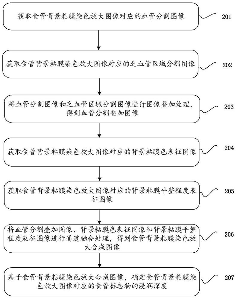
The invention provides a medical image processing method and device, a terminal and a storage medium. The method comprises the following steps of: acquiring a blood vessel segmentation image corresponding to an esophageal background mucosa staining and magnifying image; acquiring a corresponding blood-vessel-lack region segmentation image; performing image superposition processing on the blood vessel segmentation image and the blood-vessel-lack region segmentation image to obtain a blood vessel segmentation superposed image; acquiring a background mucosa color characterization image corresponding to the esophageal background mucosa staining and magnifying image; acquiring a background mucosa flatness characterization image corresponding to the esophageal background mucosa staining and magnifying image; performing channel fusion processing on the blood vessel segmentation superposed image, the background mucosa color characterization image and the background mucosa flatness characterization image to obtain an esophageal background mucosa staining and magnifying synthetic image; and determining the infiltration depth of an esophageal marker corresponding to the esophageal background mucosa staining and magnifying image based on the esophageal background mucosa staining and magnifying synthetic image. The accuracy of determining the infiltration depth of an esophageal marker is improved.

Description

technical field [0001] The present application relates to the field of medical assistance technology, and in particular to a medical image processing method, device, terminal and storage medium. Background technique [0002] With the development of gastrointestinal endoscopy technology, early diagnosis and early treatment of gastrointestinal markers and minimally invasive endoscopic treatment have been rapidly promoted. Surgical resection is the main way of diagnosis and treatment in the early stage of esophageal markers. Studies have pointed out that as the infiltration of early esophageal markers deepens, the risk of lymph node metastasis increases continuously. The risk of lymph node metastasis in physical lesions is 20-50 times higher than that in shallower lesions. Such lesions should be treated surgically, and lymph node scans should be performed if necessary. Endoscopic surgical resection has the advantages of minimal invasiveness, fewer complications, and faster rec...

Claims


Application Information

Patent Timeline
IPC IPC(8): G06T7/00G06T7/10G06T7/11
CPCG06T7/0012G06T7/10G06T7/11G06T2207/10068G06T2207/10024G06T2207/30096G06T2207/30101G06T2207/20081G06T2207/20084G06T2207/20221
Inventor 于红刚张丽辉姚理文
Owner WUHAN UNIV
Why Eureka
Try Eureka