Fundus blood vessel image segmentation method based on self-supervised learning

A supervised learning, blood vessel image technology, applied in the field of fundus blood vessel image segmentation based on self-supervised learning, can solve the problems of low contrast of eye images of blood vessel structures, inaccuracy of labeled samples, etc., and achieve the effect of improving segmentation ability and accurate pathological basis.

Pending Publication Date: 2021-12-10
CHONGQING NORMAL UNIVERSITY
View PDF0 Cites 1 Cited by
  • Summary
  • Abstract
  • Description
  • Claims
  • Application Information

AI Technical Summary

Problems solved by technology

However, due to the complexity of the vascular structure and the low contrast of eyeball images, this makes labeling samples a tedious and expensive task, and at the same time, due to the subjective factors of experts in the labeling process, it will eventually lead to inaccurate labeling samples.

Method used


Image

  • Fundus blood vessel image segmentation method based on self-supervised learning
  • Fundus blood vessel image segmentation method based on self-supervised learning
  • Fundus blood vessel image segmentation method based on self-supervised learning

Examples


Embodiment Construction

[0043] Below in conjunction with accompanying drawing, the present invention is described in further detail:

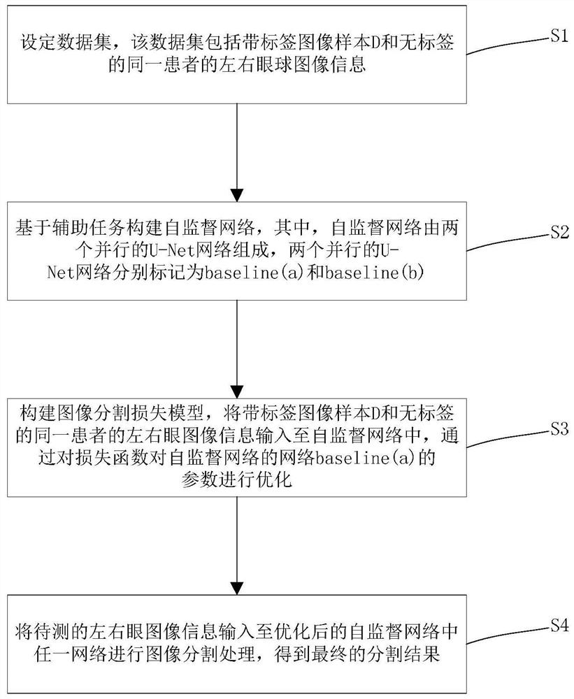
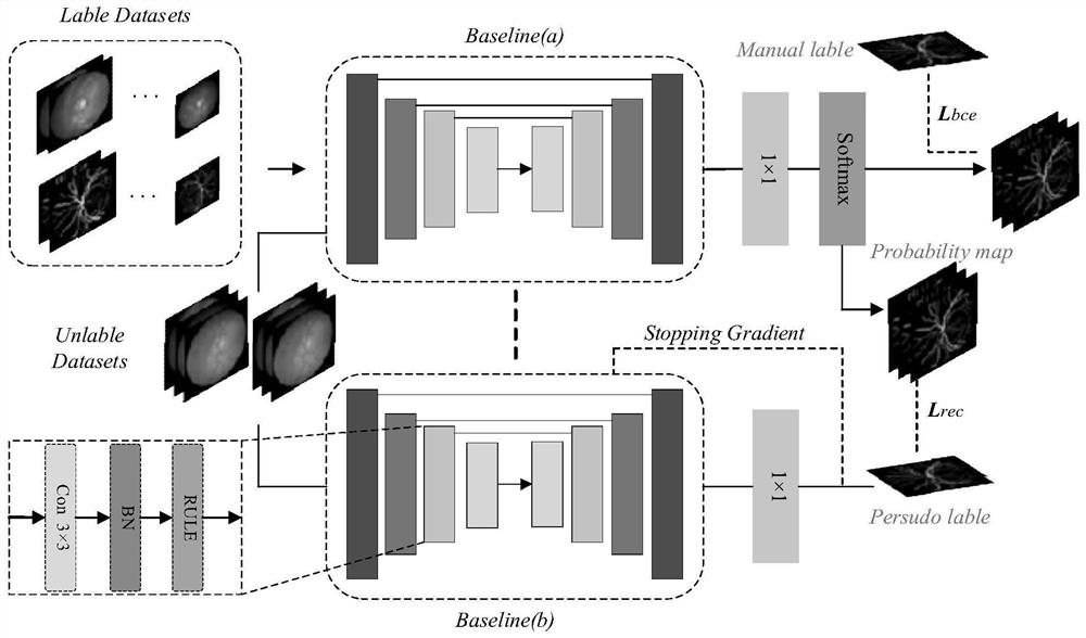
[0044] A kind of fundus blood vessel image segmentation method based on self-supervised learning provided by the present invention comprises the following steps:

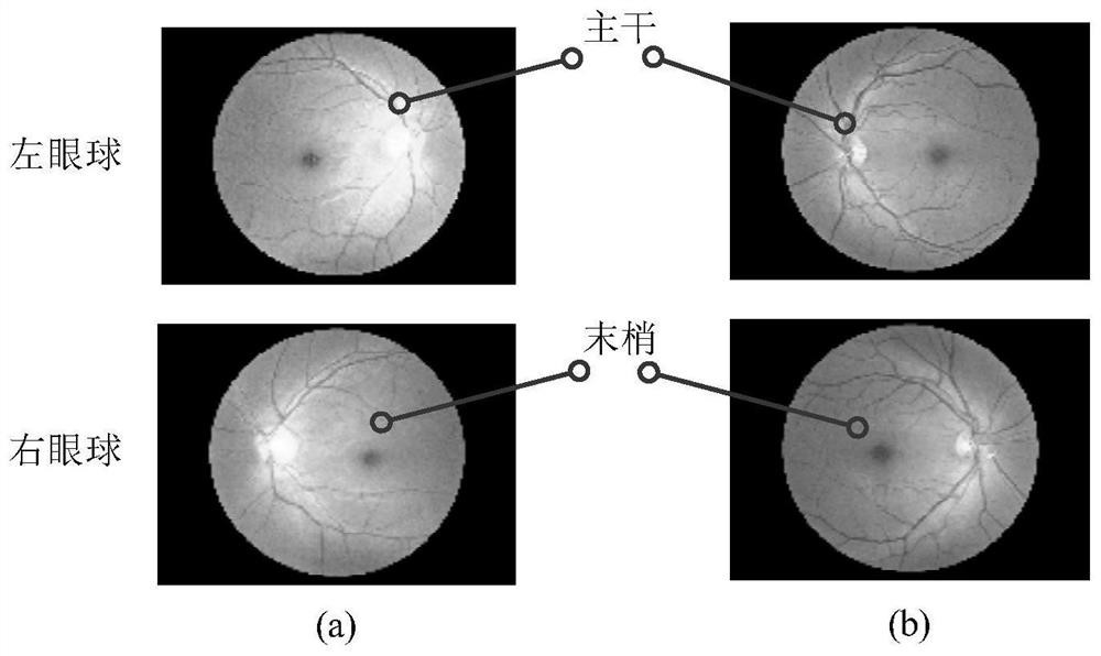
[0045] S1. Set up a data set, which includes a labeled image sample D and unlabeled left and right eyeball image information X of the same patient T∈{L,R} , where D∈R C×H×W (Y i ,X i ), where, when T=L, X L represents the left eye image of the patient, when T=R, X R represents the right eye image of the patient, R C×H×W (Y i ,X i ) represents the right-eye image of the Cth image channel, H and W represent the length and width of the image, (Y i ,X i ) represents the i-th pixel of the C-th image and the length and width of the image are H and W; among them, the labeled image sample D refers to the image after the expert segmentation process, and has segmentation label information, each image Corres...


PUM


Abstract

The invention provides a fundus blood vessel image segmentation method based on self-supervised learning. The method comprises the following steps of S1, setting a data set which comprises a labeled image sample D and label-free left and right eyeball image information XT belonging to (L, R) of a same patient; S2, constructing a self-supervised network which is composed of two identical U-Net networks based on an auxiliary task, wherein the two parallel U-Net networks are respectively marked as a baseline (a) and a baseline (b); S3, constructing an image segmentation loss model, respectively inputting the labeled image sample D and the label-free left and right eyeball image information XT belonging to (L, R) of the same patient into the baseline (a) and the self-supervision module composed of the baseline (a) and the baseline (b), optimizing the parameters of a baseline (a) network through a cross entropy loss function and a local consistency comparison loss function, wherein the network parameters of the baseline (b) in the training process are obtained through the momentum transmission of the baseline (a); and S4, inputting the to-be-detected left and right eye image information to any network in the optimized self-supervised network for image segmentation processing to obtain a final segmentation result. Through the above method, the accurate segmentation can be performed on the fundus blood vessel image.

Description

technical field [0001] The invention relates to an image segmentation method, in particular to a fundus blood vessel image segmentation method based on self-supervised learning. Background technique [0002] The image segmentation results of fundus blood vessels (that is, retinal blood vessels) can be used as the basis for the diagnosis of patients with fundus diseases, and can also provide pathological references for complications of other diseases, such as diabetes and hypertension. [0003] Among the existing fundus image segmentation algorithms, most retinal blood vessel image segmentation methods based on deep learning mostly rely on annotated images manually segmented by clinical experts. However, due to the complexity of the vascular structure and the low contrast of eyeball images, this makes labeling samples a tedious and expensive task, and at the same time, due to the subjective factors of experts in the labeling process, it will eventually lead to inaccurate labe...

Claims


Application Information

Patent Timeline
IPC IPC(8): G06T7/10G06N3/08G06K9/62G06K9/46
CPCG06T7/10G06N3/084G06T2207/20221G06T2207/20081G06T2207/30041G06F18/2415
Inventor 吕佳马超
Owner CHONGQING NORMAL UNIVERSITY
Why Eureka
Try Eureka