
Pancreatic CT image cystic tumor detection method based on attention mechanism

A CT image and detection method technology, applied in image enhancement, image analysis, image data processing, etc., can solve the problems of low detection accuracy of small pancreatic tumor lesions, missed detection and false detection, etc., achieve good detection effect and solve detection accuracy not high effect

Pending Publication Date: 2021-10-01
ZHEJIANG UNIV OF TECH
View PDF0 Cites 0 Cited by
  • Summary
  • Abstract
  • Description
  • Claims
  • Application Information

AI Technical Summary

Problems solved by technology

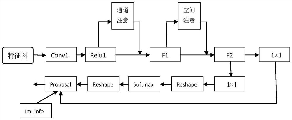
[0004] In order to solve the low detection accuracy of small pancreatic tumor lesions and the problems of missed detection and false detection, the present invention provides a method for detecting cystic tumors in pancreatic CT images based on the attention mechanism with better detection effect, combining spatial attention and channel attention force to effectively capture key areas

Method used


Image

  • Pancreatic CT image cystic tumor detection method based on attention mechanism
  • Pancreatic CT image cystic tumor detection method based on attention mechanism
  • Pancreatic CT image cystic tumor detection method based on attention mechanism

Examples


Embodiment Construction

[0016] The present invention will be further described below in conjunction with accompanying drawing:

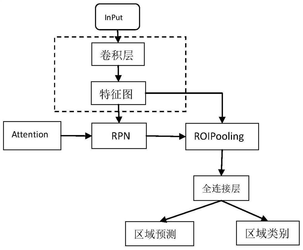
[0017] refer to Figure 1-Figure 3 , a method for detecting cystic tumors in pancreatic CT images based on an attention mechanism, comprising the following steps:

[0018] 1) Input the CT image of pancreatic cystic tumor into the network;

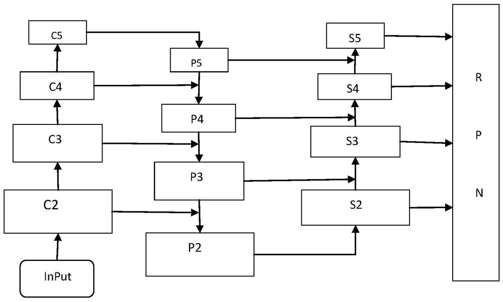
[0019] 2) Extract the features of the input image through the improved backbone based on Faster-Rcnn, and use the resnet101+FPN structure as the feature extraction network to build a bottom-up enhanced feature pyramid (such as figure 2 As shown), the input image is obtained from the bottom up through the ResNet block to obtain {C2, C3, C4, C5} feature maps of different proportions, first generate {P2, P3, P4, P5} feature maps according to FPN, and then from the P2 level Establish an augmentation path, P2 is directly used as S2 without any processing; next, in the higher resolution feature map S i A 3×3 convolution operator with strid...


PUM


Abstract

A pancreatic CT image cystic tumor detection method based on an attention mechanism comprises the steps that firstly, an overall framework is based on an improved Faster R-cnn network, and a feature extraction backbone network adopts a ResNet101 + FPN structure; a bottom-to-top enhanced feature pyramid is constructed, a multi-scale feature map generated by an FPN is input into an improved RPN network, and an attention mechanism is added into the RPN network, so that unimportant information can be effectively inhibited, and a more suitable recommendation area is generated. The problems that existing pancreatic tumor small focus detection precision is not high, and missing detection and false detection occur are solved, and the detection effect is good.

Description

technical field [0001] The invention belongs to the technical field of image target detection, and designs a convolutional neural network based on an attention mechanism for detecting cystic tumors in pancreatic CT images. Background technique [0002] Medical image object detection (MIOD) is the operation of finding objects of interest in medical images and determining their locations and categories. For example, the tumor detection of pancreatic cancer, which is a malignant tumor disease, has a 5-year survival rate of about 7%. The pancreas is a small organ located deep in the body, making detection significantly more difficult. Computed tomography (CT) compared with ultrasound and MRI, morphology is helpful in the diagnosis and staging of pancreatic cancer. Nevertheless, manual diagnosis requires doctors with extensive clinical experience, because the quality of CT images varies between different CT scanners or operators, and pathological texture features are difficult ...

Claims


Application Information

Patent Timeline
Patent Type & Authority Applications(China)
IPC IPC(8): G06T7/00G06K9/32G06K9/62G06N3/04
CPCG06T7/0012G06T2207/30096G06T2207/10081G06T2207/20081G06T2207/20084G06N3/045G06F18/2415
Inventor 管秋张跃耀苗林涛张泽涵韦子晗陈峰周乾伟
Owner ZHEJIANG UNIV OF TECH
Learn More