
Method of metal artefact reduction in x-ray dental volume tomography

A technology of tomography and X-ray, which is applied in the field of metal artifact reduction to achieve the effects of reducing processing load, reducing metal artifacts, and shortening processing time

Pending Publication Date: 2021-08-06
DENTSPLY SIRONA INC +1
View PDF1 Cites 0 Cited by
  • Summary
  • Abstract
  • Description
  • Claims
  • Application Information

AI Technical Summary

Problems solved by technology

However, this scheme is also prone to the disadvantages of the above-mentioned classical image processing techniques involving the use of thresholding

Method used


Image

  • Method of metal artefact reduction in x-ray dental volume tomography
  • Method of metal artefact reduction in x-ray dental volume tomography
  • Method of metal artefact reduction in x-ray dental volume tomography

Examples


Embodiment Construction

[0031] The reference numerals shown in the drawings refer to the elements listed below and will be referenced in the subsequent description of the exemplary embodiments.

[0032] 1.2d x-ray image

[0033] 2. Strove

[0034] 3. Patient

[0035] 3A. Patient Jaw

[0036] 4.X ray source

[0037] 5. Detector

[0038] 6. Metal object

[0039] 6A. Metal object (small volume (V) outside)

[0040] 6b. Metal object (small volume (V) inside)

[0041] 7.2D mask

[0042] 8.3D fault scan image

[0043] 9.x ray unit

[0044] V: Part of the patient's jaw (3A) or small volume

[0045] Image 6 An X-ray unit (9) is shown in accordance with an embodiment of the present invention. The X-ray unit (9) has two (V) (i.e., a small volume) for obtaining at least a portion (V) (i.e., small volume) (ie, small volume) to obtain at least a portion (V) (i.e., small volume) obtained by rotating the X-ray source (4) and detector (5) around the patient jaw (3a). Wi-X-ray image (1) or a sine chart (2) acquisition d...


PUM


Abstract

The present invention relates to a method of metal artefact reduction in x-ray dental volume tomography, the method comprising: a step (S1) of obtaining two-dimensional x- ray images (1) or a sinogram (2) of at least part (v) of a patient jaw (3a), acquired through relatively rotating an x-ray source (4) and a detector (5) around the patient jaw (3a); the method being characterized by further comprising: a step (S2) of detecting metal objects (6) in the two-dimensional x-ray images (1) or the sinogram (2) by using at least a trained artificial intelligence algorithm to generate 2D masks (7) which represent the metal objects (6) in the two-dimensional x-ray images (1) or 3D masks which represent the metal objects (6) in the sinogram (2), respectively; and a step (S4;S5) of reconstructing a three dimensional tomographic image (8) respectively based on two-dimensional x-ray images (1) or the sinogram (2) and the 2D masks (7) or the 3D masks as generated.

Description

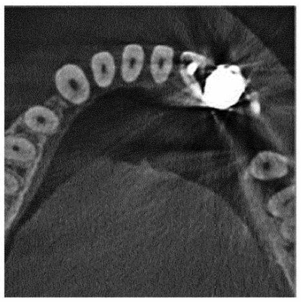
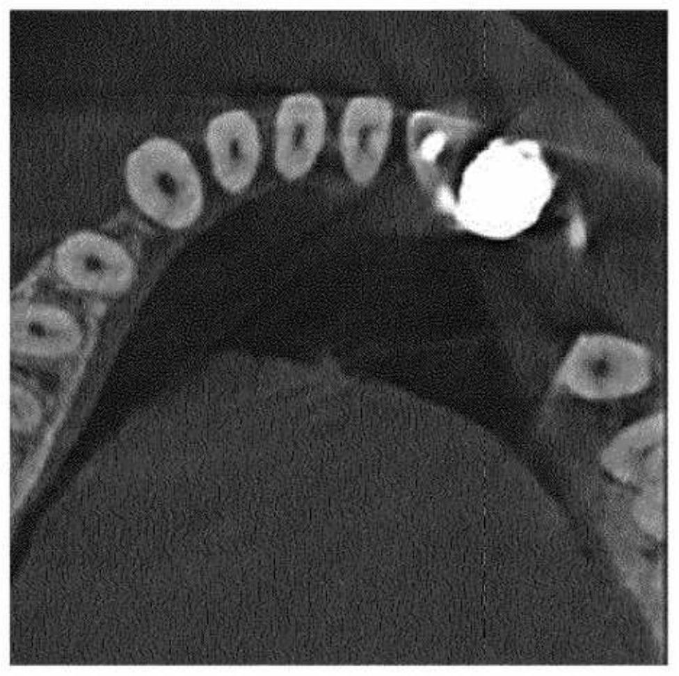
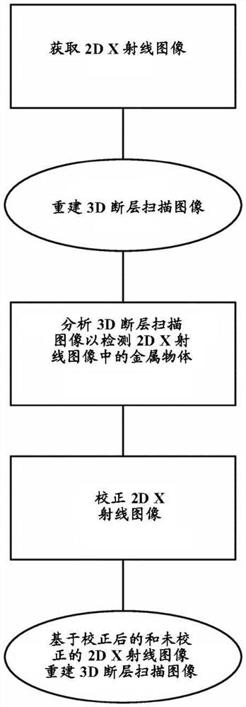
Technical field [0001] The present invention relates to X-ray dental volume breaking scanning (DVT). The invention is more particularly particularly to a method of reducing metal artifacts in an X-ray dental volume breakage. Background technique [0002] In X-ray dental volume breakdown scan, a three-dimensional (3D) tomographic scanning image of the patient's jaw is reconstructed based on two-dimensional (2D) X-ray image. Metal objects such as metal fillers or implants such as metal fillers may result in metal artifacts in the final three-dimensional fault scanning image. As shown in Figure 1, the metal artifact is displayed as dark or light stripes. These artifacts will make medical diagnosis complex. A conventional method of reducing metal artifacts is shown in FIG. In a conventional method, a two-dimensional X-ray image is first obtained, followed by reconstructing a three-dimensional (3D) tomogram scan image. Thereafter, the position of the metal object in the two-dimensiona...

Claims


Application Information

Patent Timeline
Patent Type & Authority Applications(China)
IPC IPC(8): A61B6/03A61B6/14A61B6/00G06T7/00G06T11/00A61B6/12A61B6/51
CPCA61B6/032A61B6/5258A61B6/5205A61B6/12G06T11/005G06T2211/441G06T2211/448A61B6/51G06T2210/41
Inventor C·萨尔蒂K·斯坦尼格尔M·埃尔弗斯
Owner DENTSPLY SIRONA INC
Learn More