Surgical instrument for thoracoscope
A surgical instrument and thoracoscopic technology, applied in the field of medical equipment, can solve the problem that the endoscope is easy to directly contact with human tissue, and achieve the effect of enhancing safety and reducing the degree of tissue damage
- Summary
- Abstract
- Description
- Claims
- Application Information
AI Technical Summary
Problems solved by technology
Method used
Image
Examples
Embodiment 1
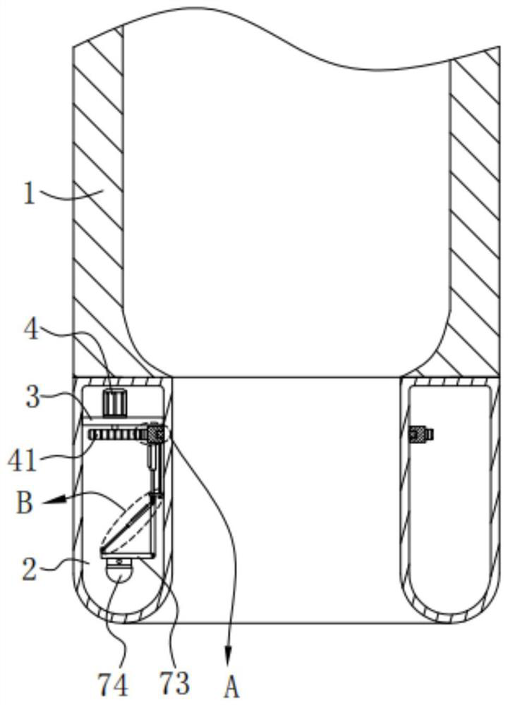
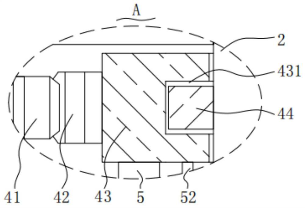
[0044] A surgical instrument for thoracoscopic surgery comprising: a connection cylinder 1 and a penetration cylinder 2, the top of the penetration cylinder 2 is fixedly connected to the bottom end of the connection cylinder 1; a fixing plate 3, the surface of the fixing plate 3 is fixed On the inner surface of the deep barrel 2; adjust the motor 4, the bottom of the adjust motor 4 is fixed on the top of the fixed plate 3, the output end of the adjust motor 4 is fixedly connected with an adjust gear 41, the adjust gear The surface of 41 is meshed with a driven gear ring 42, and the inner surface of the driven gear ring 42 is fixedly connected with a rotating ring 43; the telescopic rod 5, the top of the telescopic rod 5 is fixed on the bottom of the rotating ring 43, so The output end of the telescoping rod 5 is fixedly connected with a connection plate 51; a support frame 6, the top of the support frame 6 is fixed on the bottom of the connection plate 51, and the bottom of the...
Embodiment 2
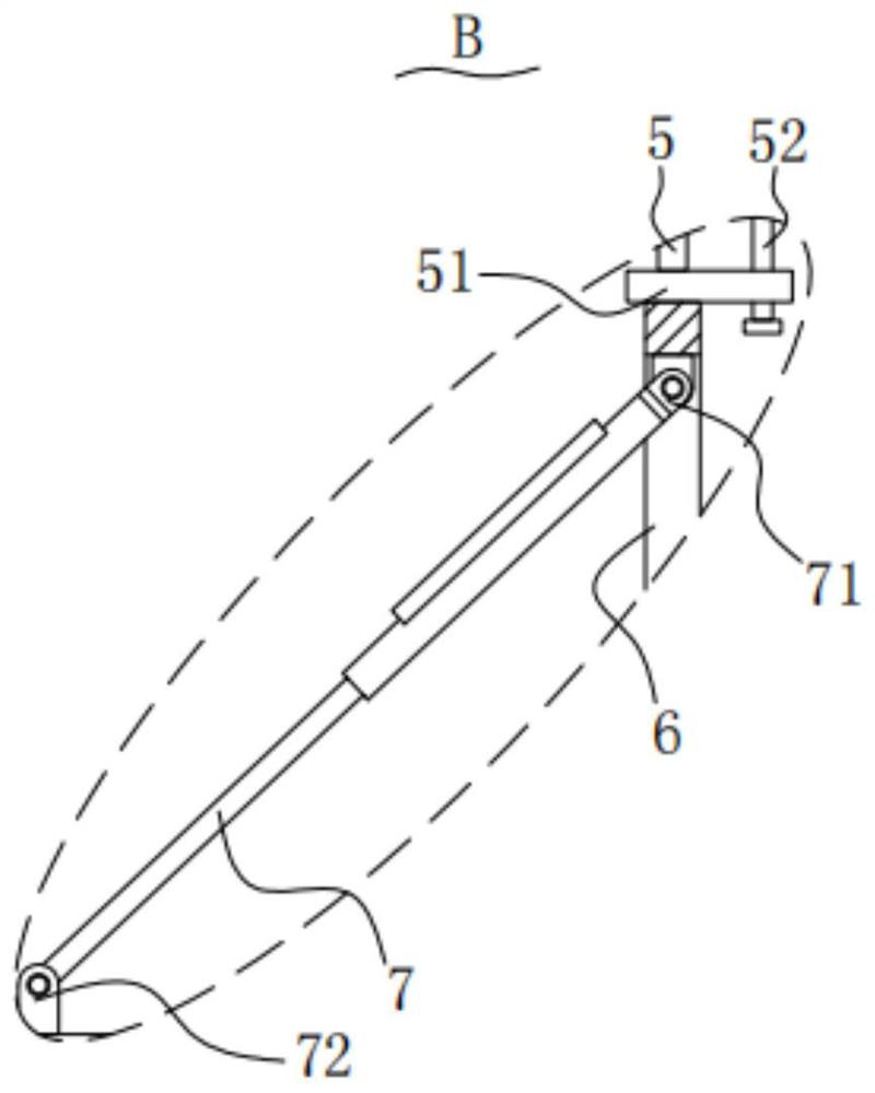
[0069] The bottom of the support frame 6 is provided with a turning rod 7 , and the two ends of the turning rod 7 are respectively provided with a first rotating member 71 and a second rotating member 72 .
[0070] The first rotating part 71 is used to connect the top end of the turning rod 7 and the inner wall of the support frame 6, and the second rotating part 72 is used to connect the surface of the mounting plate 73 and the inner wall of the turning rod 7. At the bottom end, the surface of the mounting plate 73 is rotatably connected with the inner surface of the support frame 6 through a rotating shaft.
[0071] Turnover lever 7 is an electric telescopic link, and turnover lever 7 is connected with the top of the support frame 6 inner wall through the first rotation part 71, and the output end of the turnover lever 7 is connected with the top rotation of the mounting plate 73 through the second rotation part 72, by turning over Rod 7, the first rotating part 71 and the s...
Embodiment 3
[0076] The outer surface of the penetrating cylinder 2 is covered with a protective cover film 8, and one end of the protective covering film 8 is wound from the bottom end of the penetrating cylinder 2 to the inside of the penetrating cylinder 2, for the penetrating cylinder 2 Protection during surgery.
[0077] The protective cover film 8 that goes deep into the outer surface of the tube 2 is convenient to reduce the amount of human tissue adhered to the outer wall of the deep tube 2 when the deep tube 2 is used, and improves the stability of the deep tube 2 for deep chest use.
[0078] When the penetrating tube 2 goes deep into the small hole of the thoracoscope, the human tissue is blocked on the outside by the protective cover film 8, and will not contact the outer wall of the penetrating tube 2 with a transparent structure, reducing the adhesion of tissue or effusion and affecting the inspection field of view .
[0079] The top end of the protective cover film 8 is fixe...
PUM
 Abstract
Description
Claims
Application Information
 - R&D
- Intellectual Property
- Life Sciences
- Materials
- Tech Scout
- Unparalleled Data Quality
- Higher Quality Content
- 60% Fewer Hallucinations
Browse by: Latest US Patents, China's latest patents, Technical Efficacy Thesaurus, Application Domain, Technology Topic, Popular Technical Reports.
© 2025 PatSnap. All rights reserved.Legal|Privacy policy|Modern Slavery Act Transparency Statement|Sitemap|About US| Contact US: help@patsnap.com


