Ultrasonic image blood vessel bifurcation detection method
A technology of ultrasonic image and detection method, which is applied in the field of ultrasonic image vascular bifurcation detection, to achieve the effect of removing false feature points, rich pixel points, and high efficiency
Pending Publication Date: 2021-02-12
TONGJI UNIV
View PDF6 Cites 1 Cited by
- Summary
- Abstract
- Description
- Claims
- Application Information
AI Technical Summary
Problems solved by technology
[0004] The purpose of the present invention is to provide a method for detecting blood vessel bifurcations in ultrasonic images in order to overcome the inaccurate defects in the above-mentioned prior art
Method used
View moreImage
Smart ImageViewing Examples

Examples
Embodiment
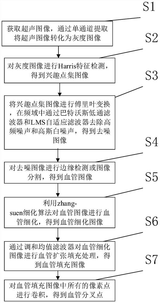
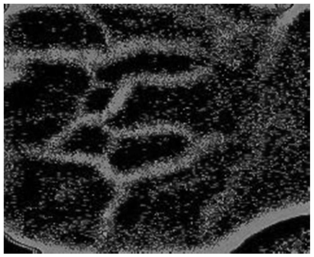
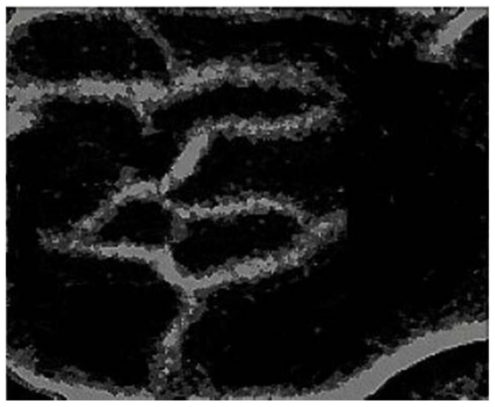
[0036] This embodiment provides a method for detecting blood vessel bifurcations in ultrasonic images, such as figure 1 shown, including the following steps:
[0037] S1: Acquire an ultrasound image, and convert the ultrasound image into a grayscale image through single-channel extraction;
Login to View More PUM
 Abstract
The invention relates to an ultrasonic image blood vessel bifurcation detection method which comprises the following steps: acquiring an ultrasonic image, and converting the ultrasonic image into a grayscale image through single-channel extraction; performing Harris feature detection on the grayscale image to obtain an interest point set image; performing Fourier transform on the interest point set image, and removing high-frequency noise and Gaussian white noise through a Butterworth low-pass filter and an LMS adaptive filter in a frequency domain to obtain a denoised image; performing edge detection or image segmentation on the denoised image to obtain a blood vessel image; carrying out blood vessel refinement on the blood vessel image by utilizing a zhang-suen refinement algorithm to obtain a blood vessel refinement image; performing blood vessel expansion filling processing on the blood vessel refined image through a harmonic mean filter to obtain a blood vessel filling image; andcarrying out convolution on all pixel points in the blood vessel filling image to obtain a blood vessel bifurcation point. Compared with the prior art, the method is simple, the determined bifurcationposition is more accurate, and the efficiency is high.
Description
technical field [0001] The invention relates to the field of detection of blood vessel bifurcations, in particular to a method for detecting blood vessel bifurcations in ultrasonic images. Background technique [0002] With the development and maturity of computer technology and the advancement of clinical diagnosis technology, medical image processing has developed rapidly. As a new discipline and technology, medical image processing technology is more and more widely used in clinical practice. At the same time, with the growth and aging of global wealth, various systemic diseases affecting the vascular network are becoming more and more common, such as age-related macular degeneration, diabetic retinopathy, glaucoma, hypertension, arteriosclerosis, and multiple sclerosis. Fundus images are often used to diagnose these lesions. Geometric features of the vasculature at bifurcations and intersections, such as intersection angles, vessel widths, bifurcation asymmetry, and ch...
Claims
Login to View More Application Information
Patent Timeline
 Patent Type & Authority Applications(China)
IPC IPC(8): G06T7/00G06T7/12G06T7/13G06T5/30
CPCG06T7/0012G06T7/13G06T7/12G06T5/30G06T2207/10132G06T2207/20056G06T2207/20081G06T2207/20104G06T2207/20221G06T2207/30101
Inventor 齐鹏侯哲林筱易葛坦谛程黎明
Owner TONGJI UNIV
Features
- R&D
- Intellectual Property
- Life Sciences
- Materials
- Tech Scout
- Unparalleled Data Quality
- Higher Quality Content
- 60% Fewer Hallucinations
Learn More Browse by: Latest US Patents, China's latest patents, Technical Efficacy Thesaurus, Application Domain, Technology Topic, Popular Technical Reports.
© 2025 PatSnap. All rights reserved.Legal|Privacy policy|Modern Slavery Act Transparency Statement|Sitemap|About US| Contact US: help@patsnap.com