Method for detecting tumor exosome-induced cell canceration process based on atomic force microscopy

An atomic force microscopy, inducing cell technology, applied in scanning probe microscopy, measuring devices, scanning probe technology, etc., can solve the problems of ignoring changes in the physical properties of exosome cells, unable to detect multi-dimensional information of cells, etc.

Pending Publication Date: 2021-01-29
CHANGCHUN UNIV OF SCI & TECH
View PDF3 Cites 0 Cited by
  • Summary
  • Abstract
  • Description
  • Claims
  • Application Information

AI Technical Summary

Problems solved by technology

[0005] However, the traditional biological method is to study the interaction between exosomes and cells from the mechanism of exosome-cell interaction, ignoring the changes of exosomes to the physical characteristics of cells, and it is impossible to intuitively detect multi-dimensional information of cells
Moreover, there is no report on the establishment of a malignant cell model induced by tumor-derived exosomes to transform benign cells into malignant cells.

Method used


Image

  • Method for detecting tumor exosome-induced cell canceration process based on atomic force microscopy
  • Method for detecting tumor exosome-induced cell canceration process based on atomic force microscopy
  • Method for detecting tumor exosome-induced cell canceration process based on atomic force microscopy

Examples


Embodiment 1

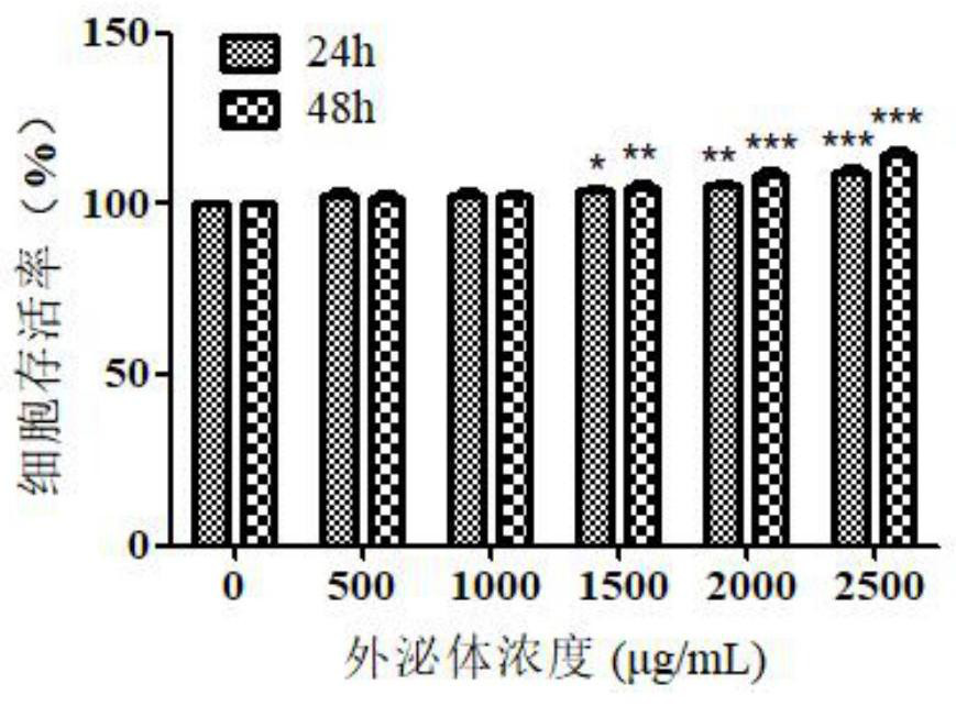
[0024] HL-7702 cells in 8×10 3 / well density into 96-well culture plates. After the cells grew to 80%, the control group was cultured with 1640 medium containing 10% FBS and 1% double antibody (penicillin / streptomycin), and the experimental group was cultured with different concentrations of HCC-LM3-exos (500, 1000, 1500, 2000 and 2500 μg / mL) culture medium. After culturing for 24h and 48h, the absorbance value of each well solution at a wavelength of 490nm was measured by the MTT method. Experimental results such as figure 1 As shown, HCC-LM3-exos at a concentration of 1500 μg / mL can significantly promote the proliferation of HL-7702 cells, and the proliferation effect is more significant with the increase of time and concentration.

Embodiment 2

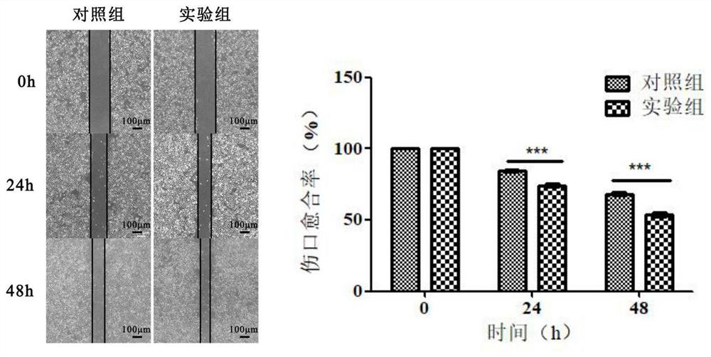
[0026] HL-7702 cells were seeded in 6-well plates. When the cell fusion rate reaches about 90% of the bottom area of ​​the culture dish, use a 200 μL yellow sterile pipette tip to draw a vertical line in the center of each well, wash with PBS twice, and remove floating cells. The experimental group was added with medium containing 1500 μg / mL HCC-LM3-exos, and the control group was cultured with normal medium. The scratched position of the cell was photographed at 0h, 24h and 48h respectively. Such as figure 2 As shown, the results showed that the wound healing rate of the cells in the experimental group was significantly higher than that in the control group, and the migration effect was more significant with the increase of time.

Embodiment 3

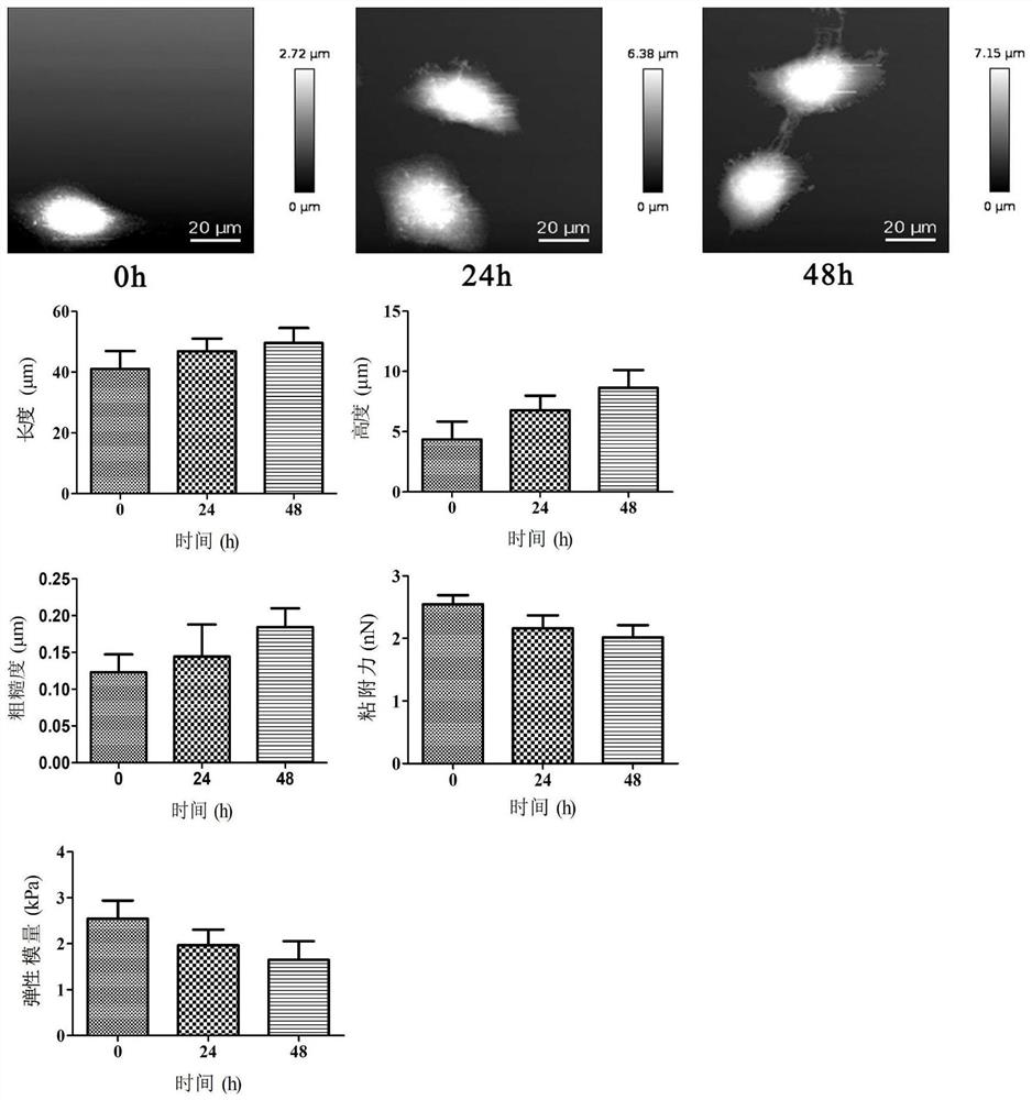
[0028] The morphology, elastic modulus, adhesion, and surface roughness of cells were measured using an atomic force nanomanipulation system. First, HL-7702 cells were seeded on coverslips at an appropriate density at 37 °C, 5% CO 2 cultured under concentration conditions. The control group was cultured with normal medium, and the experimental group was added with HCC-LM3-exos at a concentration of 1500 μg / mL. After culturing for 24h and 48h, the cells were detected. The model of the atomic force probe used in the experiment is SHOCONG-10, and the spring constant used in the experiment is 0.1N / m. Experimental results such as image 3 As shown, the edges of the cells in the control group were clear, but after being treated with HCC-LM3-exos for different times, the edges of the cells were blurred and showed a state of outward diffusion. As the treatment time of HCC-LM3-exos increased, the cell length, height, and surface roughness increased, while the adhesion force and ela...


PUM


Abstract

The invention provides a method for detecting tumor-derived exosome-induced cell canceration based on atomic force microscopy, and belongs to the technical field of atomic force microscopy imaging. The exosome is obtained by treatment through an ultrafiltration tube centrifugation method, tumor-derived exosome and normal cells are co-cultured to obtain a malignant cell model, the proliferation andmigration capabilities of cells are detected, and meanwhile, multidimensional information detection is performed on the cells through atomic force microscopy. According to the detection method provided by the invention, real-time imaging, quantitative monitoring and visual operation can be carried out on a malignant cell model for benign cell transformation in a liquid environment, so that the overall form, elasticity modulus, adhesive force, surface roughness and membrane potential (mechanical property and electrical property) change of cells can be obtained; the multi-dimensional information detection of the cell malignant evolution dynamic process is realized, and the technical support is provided for the research of cell heterogeneity and cell vicious change new theories.

Description

technical field [0001] The invention relates to the technical field of atomic force microscopy imaging, in particular to a method for detecting malignant transformation of cells induced by tumor-derived exosomes based on atomic force microscopy. Background technique [0002] Exosomes are tiny vesicles with a lipid bilayer membrane structure that are secreted by cells and are currently believed to be about 30-150 nm in diameter. Exosomes are rich in nucleic acids, proteins, and lipids, and are widely distributed in body fluids such as blood, saliva, and urine. As a medium for intercellular communication, exosomes have a variety of biological functions. In recent years, multiple studies have shown that the secretion and dysfunction of exosomes play a crucial role in the occurrence, development and treatment of malignant tumors. [0003] Exosomes secreted by tumor cells (called tumor-derived exosomes), studies have shown that tumor-derived exosomes enhance the invasion and mig...

Claims


Application Information

Patent Timeline
Patent Type & Authority Applications(China)
IPC IPC(8): G01Q60/24
CPCG01Q60/24
Inventor 王作斌鞠拓宇王树伟王佳佳陈玉娟杨帆姜晓琳董莉彤宋正勋许红梅张景然
Owner CHANGCHUN UNIV OF SCI & TECH
Why Eureka
Try Eureka