Multicolor fluorescence microscopic imaging system

A microscopic imaging and fluorescence technology, applied in the field of multi-color fluorescence microscopy imaging systems, can solve the problems of low spatial resolution, poor signal-to-noise ratio, low quantum efficiency, etc., and achieve improved information dimension, high integration and stability. , the effect of high temporal resolution

Pending Publication Date: 2022-05-06
CHANGCHUN INST OF OPTICS FINE MECHANICS & PHYSICS CHINESE ACAD OF SCI
View PDF0 Cites 0 Cited by
  • Summary
  • Abstract
  • Description
  • Claims
  • Application Information

AI Technical Summary

Problems solved by technology

Due to the low quantum efficiency, low spatial resolution, and poor signal-to-noise ratio of existing color detectors, biological multispectral imaging mainly uses spectral separation to convert spectral information into spatial intensity distribution, and monochromatic detectors are used for multispectral imaging. spectral imaging
There are three main ways to achieve spectral separation: sequential switching of emission filters, prism or grating dispersion, and sequential dichroic beam splitting. These three spectral separation methods are respectively in sampling synchronization, spectral resolution, and device complexity. obvious flaws in

Method used


Image

  • Multicolor fluorescence microscopic imaging system
  • Multicolor fluorescence microscopic imaging system
  • Multicolor fluorescence microscopic imaging system

Examples


Embodiment Construction

[0025] Hereinafter, embodiments of the present invention will be described with reference to the accompanying drawings. In the following description, the same blocks are denoted by the same reference numerals. With the same reference numerals, their names and functions are also the same. Therefore, its detailed description will not be repeated.

[0026] In order to make the object, technical solution and advantages of the present invention clearer, the present invention will be further described in detail below in conjunction with the accompanying drawings and specific embodiments. It should be understood that the specific embodiments described here are only used to explain the present invention, but not to limit the present invention.

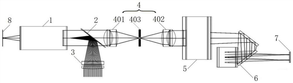
[0027] figure 1 The structure of the multi-color fluorescence microscopic imaging system provided according to the embodiment of the present invention is shown.

[0028] Such as figure 1 As shown, the multicolor fluorescence microscopy im...


PUM


Abstract

The invention provides a multicolor fluorescence microscopic imaging system. The multicolor fluorescence microscopic imaging system comprises an objective lens, a dichroscope, a convergent mirror, an achromatic relay lens group, a barrel lens, a beam splitter and a detector, wherein the exciting light is focused on an object to be detected through the convergent mirror and the dichroscope to excite fluorescence, and the fluorescence is collected through the objective lens, then is converged through the achromatic relay lens group and the barrel lens, enters the beam splitter to be subjected to beam splitting and is respectively imaged on the detector. On the basis of not increasing hardware required by the imaging system such as a detector and an objective lens, the imaging system is formed in a relatively simple mode, so that the information dimension in single acquisition or unit time is improved, and meanwhile, the imaging mode also has extremely high time resolution.

Description

technical field [0001] The invention relates to the technical field of multicolor microscopic imaging, in particular to a multicolor fluorescent microscopic imaging system. Background technique [0002] Multicolor fluorescence microscopy imaging utilizes the specific biological binding and selective stimulated emission spectra of fluorescent probes to obtain extremely high signal-to-noise ratio and spatial resolution, especially in biological microstructure imaging and high-throughput It has important applications in the field of bioinformatics detection. Due to the low quantum efficiency, low spatial resolution, and poor signal-to-noise ratio of existing color detectors, biological multispectral imaging mainly uses spectral separation to convert spectral information into spatial intensity distribution, and monochromatic detectors are used for multispectral imaging. spectral imaging. There are three main ways to achieve spectral separation: sequential switching of emission...

Claims


Application Information

Patent Timeline
Patent Type & Authority Applications(China)
IPC IPC(8): G01N21/64G01N21/01
CPCG01N21/6458G01N21/6486G01N21/6428G01N21/01G01N2021/0112
Inventor 吴一辉高明王越
Owner CHANGCHUN INST OF OPTICS FINE MECHANICS & PHYSICS CHINESE ACAD OF SCI
Why Eureka
Try Eureka