Method for transfecting cells through ultrasonic perforation

A technology of transfection cells and ultrasound, which is applied in the field of cell transfection, can solve the problems of high cell death rate, virus risk restricting large-scale use, cell damage, etc., and achieves simple operation, reduced professional skill requirements and time investment requirements , the effect of cost reduction

Active Publication Date: 2021-01-26
SUZHOU INST OF BIOMEDICAL ENG & TECH CHINESE ACADEMY OF SCI
View PDF9 Cites 2 Cited by
  • Summary
  • Abstract
  • Description
  • Claims
  • Application Information

AI Technical Summary

Problems solved by technology

Moreover, the risk of the virus to cells or in the body also limits its large-scale use
In the non-viral transfection system, electroporation transfection is the most widely used method, but the high voltage during the electroporation process and the large amount of hydroxide ions generated by the cathodic effect cause great damage to the cells, resulting in cells after electroporation. high death rate
At the same time, the transfection of a small number of cells (microliters to tens of milliliters) targeted by the current electroporation infection equipment is powerless for the transfection of pharmaceutical companies that need to expand the scale to hundreds of milliliters

Method used


Image

  • Method for transfecting cells through ultrasonic perforation
  • Method for transfecting cells through ultrasonic perforation
  • Method for transfecting cells through ultrasonic perforation

Examples


Embodiment 1

[0036] Example 1 Ultrasound-mediated gene transfection

[0037] A method for ultrasonically perforating and transfecting cells, comprising the following steps:

[0038] 1) Culture Hela cells (target cells):

[0039] Cultivate Hela cells in a 6-well plate, and cultivate them for 3 days at 37°C in a 5% carbon dioxide atmosphere, and the culture medium is RPMI medium (Gibco company) containing 2.5% bovine serum (sigma company); wash the cells twice with PBS Finally, trypsin-EDTA was added to digest the cells for 2 minutes, and then RPMI medium containing 2.5% bovine serum was added; after the cells were gently blown down to form a single cell suspension, centrifuged at 1200 rpm for 5 minutes at room temperature.

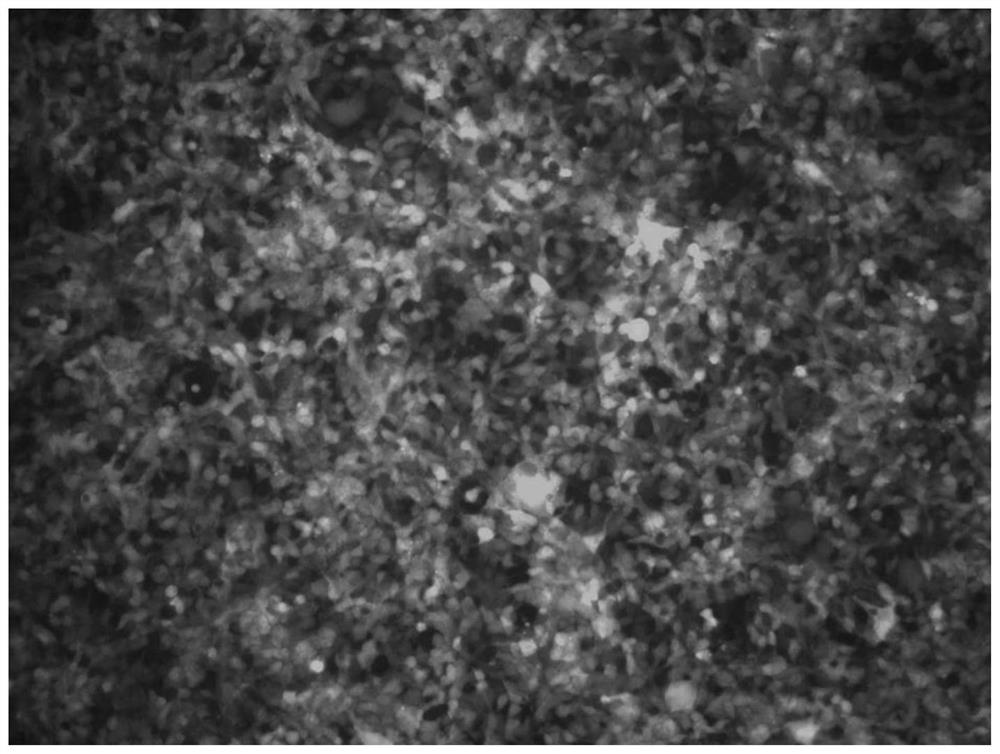
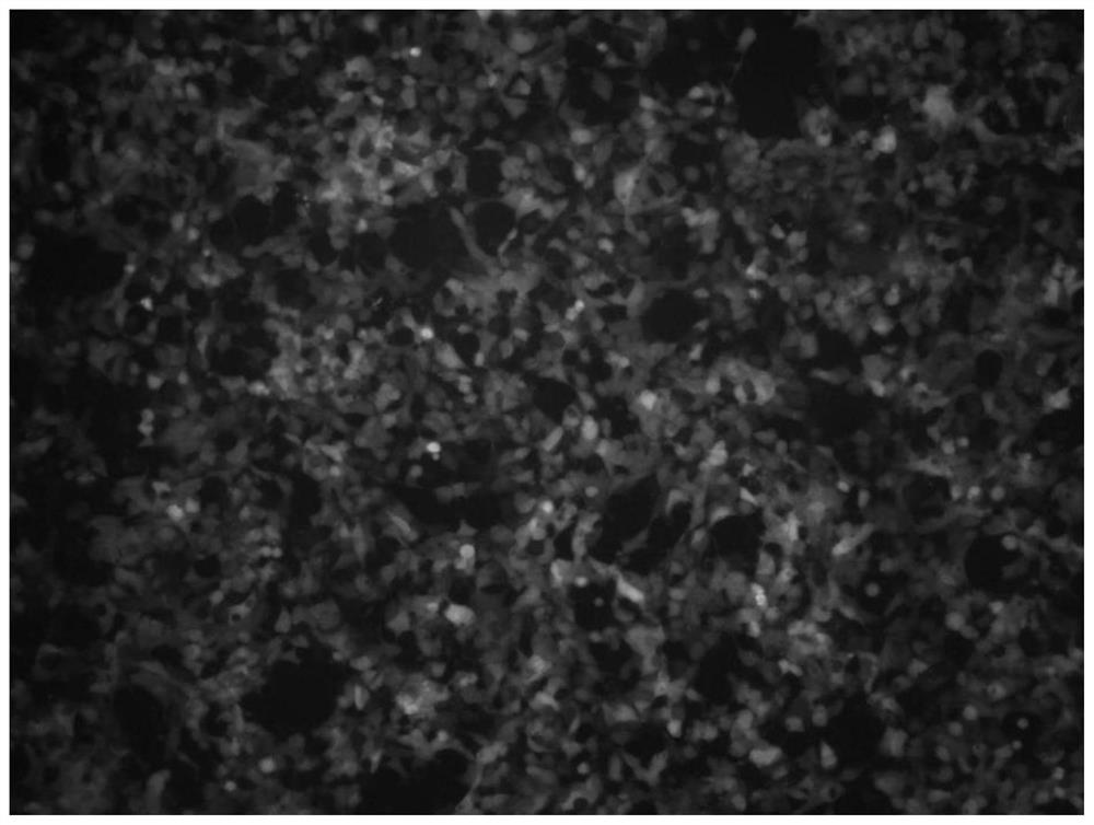
[0040] 2) Configure the ultrasound contrast agent (SonoVue, Bracco company) in 5ml PBS buffer, then add target cells, pEGFP-C1 plasmid (target gene) with green fluorescence, wherein the final concentration of the target gene is 20ug / ml , the final concentration of ult...

Embodiment 2

[0043] Example 2 Ultrasound-mediated protein transfection

[0044] A method for ultrasonically perforating and transfecting cells, comprising the following steps:

[0045] 1) cultivating Hela cells (target cells), the same as the method in Example 1;

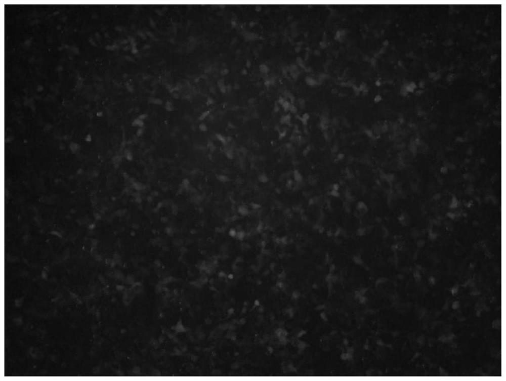
[0046]2) Resuspend 1ug of target protein: fluorescently labeled bovine serum albumin (FITC-BSA, purchased from Sigma) in 10ul ultrasound contrast agent to obtain a mixture, then take 2.5ul of the mixture and mix it with 200ul target cells in In the glass tube and then mixed with the target cells in the glass tube;

[0047] 3) Place the transducer used to emit ultrasonic waves at the bottom of the glass tube, and keep the distance between the transducer and the bottom of the glass tube at 10-30mm, adjust the frequency of ultrasonic waves to 800-1200kHz, so that the transducer emits Ultrasound acts on the glass tube for 30-120s; refer to Figure 4 , adopt ultrasonic generator to produce ultrasonic wave in the present embodiment...


PUM


Abstract

The invention discloses a method for transfecting cells through ultrasonic perforation. The method comprises the following steps: firstly, culturing target cells; secondly, mixing the target cells, atarget object and an ultrasonic contrast agent in a container; thirdly, applying ultrasonic waves to the container; and fourthly, re-culturing the container under a constant temperature condition, sothat the target object fully enters the target cells, wherein the target object is a target gene or a target protein, and when the target object is the target gene, the frequency of the ultrasonic waves adopted in the third step is 800-900 kHz; and when the target object is the target protein, the frequency of the ultrasonic waves adopted in the third step is 800-1200 kHz. The method for transfecting cells through ultrasonic perforation is simple to operate, high in transfection efficiency and low in cost, and can be used for converting or transfecting molecules such as DNA (Deoxyribonucleic Acid), RNA (Ribose Nucleic Acid), proteins, viruses, sugar, medicines and nanoparticles in an unlimited range.

Description

technical field [0001] The invention relates to the technical field of cell transfection, in particular to a method for ultrasonically transfecting cells. Background technique [0002] Cell engineering, genetic engineering, genetic engineering and other disciplines or industries all involve eukaryotic cells (such as bacteria) and prokaryotic cells (such as fungi, mice, sheep, human cells), etc. Transport of viruses, proteins, drugs, nanomaterials, etc. from extracellular to intracellular. In the field of life science research, this process is a necessary process for cells to obtain new genetic traits or protein expression; in the development of new drugs, this process is applied to small batch protein / antibody preparation, antibody drug screening, and positive clone screening; In immunotherapy, it is also a key technical link in the transient preparation of CAR-T, CAR-NK and other cells. Therefore, the transport of molecules into cells involves the whole industry of life s...

Claims


Application Information

Patent Timeline
Patent Type & Authority Applications(China)
IPC IPC(8): C12N15/85C12N13/00
CPCC12N15/85C12N13/00C12N2800/107
Inventor 宋一之郄兴旺马玉婷王策
Owner SUZHOU INST OF BIOMEDICAL ENG & TECH CHINESE ACADEMY OF SCI
Why Eureka
Try Eureka