
Brain tumor segmentation method of deep residual network based on U-Net architecture.

A brain tumor and network technology, applied in the field of medical image segmentation and deep learning, can solve the problem of not fully considering the important role of residual learning, and achieve the effect of preventing network performance degradation, advanced performance, and improving network performance.

Active Publication Date: 2021-01-08
JINING UNIV
View PDF6 Cites 2 Cited by
  • Summary
  • Abstract
  • Description
  • Claims
  • Application Information

AI Technical Summary

Problems solved by technology

Both of these methods and the standard U-net method do not fully consider the important role of residual learning in deep learning networks

Method used


Image

  • Brain tumor segmentation method of deep residual network based on U-Net architecture.
  • Brain tumor segmentation method of deep residual network based on U-Net architecture.
  • Brain tumor segmentation method of deep residual network based on U-Net architecture.

Examples


Embodiment Construction

[0055] In order to make the objectives, technical solutions and advantages of the present invention more clearly understood, the present invention will be further described in detail below according to the accompanying drawings and examples.

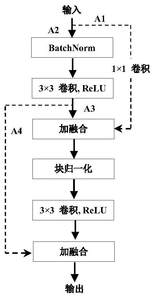
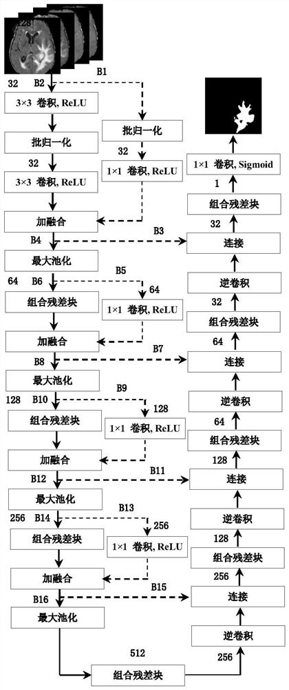
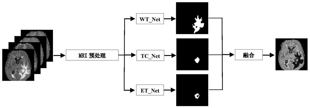
[0056] The brain tumor segmentation process proposed by the present invention is a learning algorithm based on a convolutional neural network. The framework of the method consists of the following three main steps: 3D MRI data preprocessing, neural network training, and application of the trained algorithmic model to predict the structure of brain tumors.

[0057] A. Preprocessing

[0058] Before segmentation, it is usually necessary to preprocess the segmented brain images to eliminate the influence of irrelevant information, thereby improving the reliability of image segmentation results. In fact, even though MRIs from the same patient are all collected on the same scanner, MRIs can vary at different times or under pathological condit...


PUM


Abstract

The invention discloses a brain tumor segmentation method of a deep residual network based on U-Net architecture. The brain tumor segmentation method comprises the following steps: S1, constructing acombined convolution block of the deep residual network; and S2, constructing a branch structure network for implementing the three-branch structure model; and S3, combining to form an integral network of the three-branch structure model. The method has the advantages that the network performance can be more effectively prevented from being reduced, and the network performance can be better improved. Besides, standardizing, cutting and removing the focus-free image data of each modal image data, and finally combining the focus-free image data into multi-channel data. Therefore, experimental results show that the algorithm has advanced performance. Tumor segmentation effect is greatly improved.

Description

technical field [0001] The invention relates to the technical field of medical image segmentation and deep learning, in particular to a brain tumor segmentation method based on a deep residual network of U-Net architecture. Background technique [0002] Quantitative analysis of magnetic resonance images (MRI) of the brain has become routine for many neurological disorders and is the method of choice for structural brain analysis due to the high contrast and high spatial resolution of such images to soft tissues without known health risk. The basic principle of MRI is that each hydrogen proton in the human body can be regarded as a small magnet, and the signals generated by the hydrogen nuclei in the magnetic field are processed by a computer to reconstruct the image. MRI signals can accurately display the anatomical structure of the central nervous system, the location of tumors and other lesions, the size of the area, and the relative positional relationship with other sur...

Claims


Application Information

Patent Timeline
Patent Type & Authority Applications(China)
IPC IPC(8): G06T7/00G06T7/11G06T7/194G06K9/62G06N3/04G06N3/08
CPCG06T7/0012G06T7/11G06T7/194G06N3/084G06T2207/10088G06T2207/20081G06T2207/20084G06T2207/30016G06T2207/30096G06V2201/03G06N3/045G06F18/241G06F18/253
Inventor 黄传波刘传领丁华立
Owner JINING UNIV
Learn More