
Human extracellular matrix protein 1 detection kit

A kit and protein technology, applied in the field of diagnosis, can solve problems such as failure to detect liver fibrosis, failure to reflect the whole, patient death, etc.

Pending Publication Date: 2020-12-11
INST PASTEUR OF SHANGHAI CHINESE ACADEMY OF SCI
View PDF1 Cites 0 Cited by
  • Summary
  • Abstract
  • Description
  • Claims
  • Application Information

AI Technical Summary

Problems solved by technology

[0003] The existing diagnosis of liver fibrosis is mainly divided into the following categories: 1. Liver biopsy pathological examination is still the gold standard for diagnosing liver fibrosis and cirrhosis at present, but there are still some cases such as death of patients, partial failure to reflect the whole, etc. shortcoming
2. Non-invasive testing such as Fibroscan
Fibroscan often needs to be tested jointly with other indicators, and due to the influence of ascites and fat deformation of the patient, the accuracy of the test results is slightly lacking
3. Serological testing mainly includes a series of indicators such as serum type Ⅲ procollagen amino terminal peptide, serum laminin, serum hyaluronic acid, serum collagen Ⅳ, matrix metalloproteinase, etc. The existing serological indicators are also specific and accurate Therefore, it cannot be an excellent indicator for the detection of liver fibrosis
The reasons are as follows: 1) Most of the direct serological indicators reflect the metabolic rate of the substrate and are closely related to the inflammatory response. When there is inflammation in the body, the degree of liver fibrosis cannot be specifically detected.
2) Most of them are not liver-specific

Method used


Image

  • Human extracellular matrix protein 1 detection kit
  • Human extracellular matrix protein 1 detection kit
  • Human extracellular matrix protein 1 detection kit

Examples


Embodiment 1

[0233] Example 1 Obtaining of human ECM1 gene

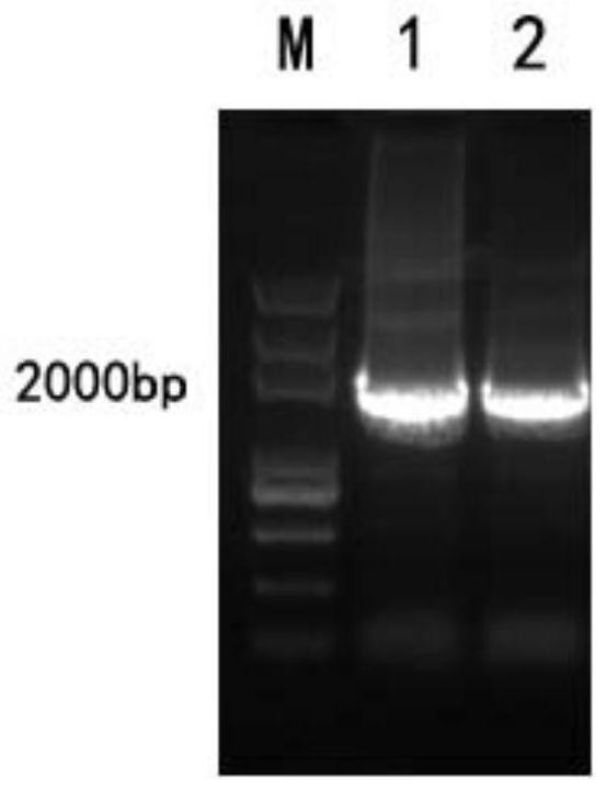
[0234] Using the THp1 cells preserved in our laboratory as a template, a fragment of about 1900bp was amplified with Human-ECM1-sense and Human-ECM1-anti primers, and the PCR product was recovered by running rubber and tapping, and then digested with NotI and BamHI double enzymes, and connected to the same On the pcDNA3.1-FC expression vector treated with double enzyme digestion, transform Escherichia coli DH5α competent, the plasmid insert fragment was verified by bacterial liquid PCR, and the positive clone was selected for DNA sequencing (sequencing was completed by Bosun Biological Co., Ltd.) after the reagents were correct. The plasmid was extracted using the cassette to obtain the expression plasmid pcDNA3.1-Fc-hECM1.

[0235] From figure 1 It can be seen from the figure that the human ECM1 gene was successfully obtained with good purity and content.

Embodiment 2

[0236] The preparation of embodiment 2 monoclonal antibody

[0237] 2.1 Monoclonal Antibody Protein G Affinity Chromatography Purification

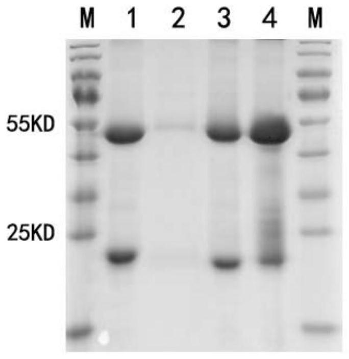
[0238] Inject 0.5 mL pristane intraperitoneally into the mice. After 7-10 days, intraperitoneally inject 0.5mL 1×10 6 hybridoma cells. The growth of the mice was observed every day, and abdominal swelling was visible in about 7 days, and the ascites was collected in time. The ascites fluid collected from 4 mice was preliminarily purified by 50% saturated ammonium sulfate precipitation, and then purified by Protein G-sepHarose4B. The protein not bound to the column was eluted by PB, and the protein was finally eluted by 0.1mol / L pH2.7glycine. After protein G was collected and purified, the antibody was added to the loading buffer and boiled, loaded, electrophoresed by SDS, and stained with Coomassie brilliant blue. Check the effect of antibody purification.

[0239] Experimental results such as figure 2 As shown, the four antibodies...

Embodiment 3

[0242] Example 3 Characterization of Anti-hECM1 monoclonal antibody after purification

[0243] The amount of biotin reagent added was calculated according to the concentration of the purified antibody, and the 4 murine monoclonal antibodies were labeled with Biotin and purified through a desalting column. Then, the affinity of the labeled monoclonal antibody to the standard was detected by indirect ELISA, and the standard was serially diluted (10ng / mL, 5ng / mL, 2.5ng / mL, 1.25ng / mL, 0.625ng / mL, 0.31ng / mL mL, 0.15ng / mL, 0.075ng / mL). The affinities of the Biotin-labeled antibodies to the standards with different concentrations were tested respectively.

[0244] Experimental results such as Figure 4 As shown, the OD value of the purified and Biotin-labeled antibody was much higher than that of the negative control group, indicating that the affinity activity of the purified and labeled antibody was not affected by purification and Biotin labeling, and the No. 4 and No. 3 antibo...


PUM

PropertyMeasurementUnit
Sensitivityaaaaaaaaaa

Abstract

The invention relates to a human extracellular matrix protein 1 (ECM1) detection kit. Specifically, the invention provides a monoclonal antibody, a reagent and a kit for detecting an ECM1 protein. Theantibody is produced by a hybridoma cell line CCTCC NO:C2019109 or CCTCC NO:C2019110. The reagent or the kit provided by the invention can be used for detecting a quantity of the human ECM1 protein with extremely low concentration and reflecting the degree of human hepatic fibrosis, and is very high in sensitivity and accuracy, good in repeatability, simple, convenient and rapid to operate, low in cost, non-invasive and suitable for rapid quantitative detection.

Description

technical field [0001] The present invention relates to the field of diagnostics. More specifically, the present invention relates to a human extracellular matrix protein 1 (ECM1) detection kit. Background technique [0002] Since liver fibrosis is reversible and liver cirrhosis is an irreversible process, safe and non-invasive early diagnosis plays a very important role in the treatment of liver fibrosis. [0003] The existing diagnosis of liver fibrosis is mainly divided into the following categories: 1. Liver biopsy pathological examination is still the gold standard for diagnosing liver fibrosis and cirrhosis at present, but there are still some cases such as death of patients, partial failure to reflect the whole, etc. shortcoming. 2. Fibroscan and other non-invasive testing. Fibroscan reflects the liver parenchyma stiffness by measuring the instantaneous liver elasticity map, judges the degree of liver fibrosis by measuring liver stiffness, and accurately grades liv...

Claims


Application Information

Patent Timeline
IPC IPC(8): G01N33/68G01N33/577
CPCG01N33/577G01N33/6803G01N2800/085
Inventor 孙兵李楠范卫国伊春艳凌志洋
Owner INST PASTEUR OF SHANGHAI CHINESE ACADEMY OF SCI
Learn More