
Animal magnetic resonance imaging eye protection device and use method

A technology of magnetic resonance imaging and protective devices, applied in applications, medical science, sensors, etc., can solve problems such as difficult to protect animals, affecting animals, unfavorable protective measures, etc.

Pending Publication Date: 2020-10-23
HARBIN MEDICAL UNIVERSITY
View PDF8 Cites 1 Cited by
  • Summary
  • Abstract
  • Description
  • Claims
  • Application Information

AI Technical Summary

Problems solved by technology

However, in order to observe the occurrence and development of the disease, the modeling process of model animals requires repeated MRI scans at different time points. If the animal's eyes are sick or blind, it will affect the animal's diet, mood, etc., which is not conducive to specific conditions. Successful establishment of disease models and further long-term follow-up studies
[0004] Animals should take measures to protect their eyes during the MRI process, but the animals are in the center of a strong magnetic field, which is not conducive to the use of conventional protective measures
If you wait for one inspection to be completed, exit the scanning bed, apply protective measures, and then proceed to the next scan, the entry and exit of the scanning table need to be repositioned, which not only affects the experimental process, but also makes it difficult to ensure the layer position and direction of the same inspection site before and after the animal. Consistent, not conducive to later image comparison
[0005] At present, the risk of blindness in the eyes of animals is basically not considered or measures are taken when performing magnetic resonance imaging experiments on animals, but the protective measures are complicated and cover the entire head area, which is not conducive to head-related inspections

Method used


Image

  • Animal magnetic resonance imaging eye protection device and use method
  • Animal magnetic resonance imaging eye protection device and use method
  • Animal magnetic resonance imaging eye protection device and use method

Examples


Embodiment 1

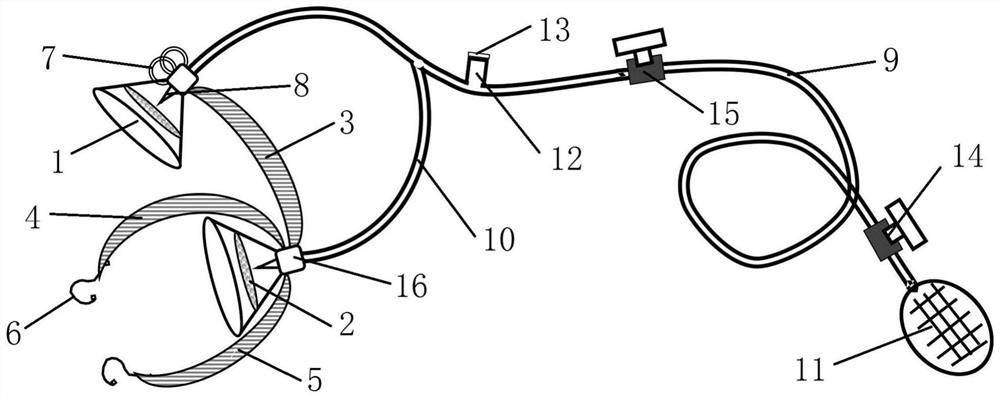
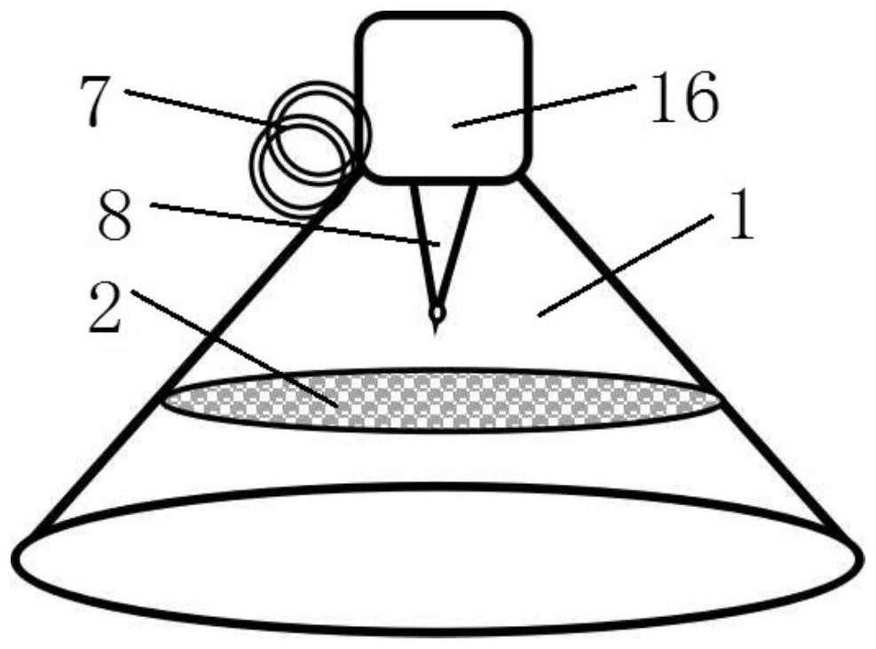
[0030] Such as Figure 1-2 As shown, an animal magnetic resonance imaging eye protection device includes two conical eye masks 1, the conical eye masks 1 are conical hard non-magnetic plastic structures, and a layer of cotton gauze 2 is arranged inside the conical eye masks 1, cotton gauze 2 The distance between the quality gauze 2 and the open end of the conical eye mask 1 is 3 mm, so as to prevent the liquid from directly dripping on the eyes of the animal, which will irritate both eyes and cause discomfort and local movement of the eyes of the animal.

[0031] The two conical eye masks 1 are provided with a connection mechanism fixed to the head of the animal. The connection mechanism includes a head rear strap 3, a head front strap 4 and a chin strap 5, and the head rear strap 3, the head front strap 4 and the chin strap 5 are all Elastic fixing band, the two ends of band 3 behind the head link to each other with two conical eye masks 1 respectively, one end of band 4 and ...

Embodiment 2

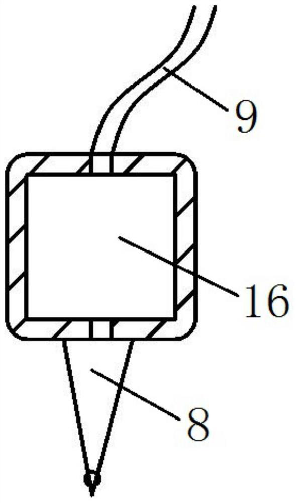
[0035] This embodiment is basically the same as Embodiment 1, the difference is: as figure 1 with 3 As shown, the top of the conical eye mask 1 is sleeved with a liquid storage box 16, the liquid outlet of the second delivery pipe is connected with the upper end of the liquid storage box 16, and the liquid inlet of the pressure nozzle 8 is connected with the lower end of the liquid storage box 16. The second delivery pipe communicates with the pressure nozzle 8 through the liquid storage box 16 . The two ends of the headband 3 are respectively connected to the side walls of the corresponding liquid storage box 16, and one end of the head front belt 4 and the chin belt 5 is connected to the side wall of a liquid storage box 16, and the two hanging rings 7 are fixed on the other side. On the side wall of a liquid storage box 16. The liquid storage box 16 is added to increase the storage capacity of the liquid medicine in the air delivery assembly, and at the same time, it is c...

Embodiment 3

[0037] A method for using the animal magnetic resonance imaging eye protection device described in Embodiment 1 and Embodiment 2, comprising the following steps:

[0038] (1) Before the magnetic resonance scan starts, inject physiological saline or eye drops into the first air delivery pipe 9 and the second air delivery pipe 10 through the water injection port 12 of the first air delivery pipe 9 and seal the water injection port 12;

[0039] (2) Fix the experimental animal on the MRI scanning bed, and then cover the eyes of the animal with two conical goggles 1 respectively. The hanging ring 7 is connected, and the chin strap 5 is located at the chin of the animal and connected with another hanging ring 7, so that the conical eye mask 1 is fixed on the head of the animal;

[0040] (3) Open the first one-way valve 14 and the second one-way valve 15, and manually press the hand-held airbag, the gas enters the first air delivery pipe 9 through the first one-way valve 14, the firs...


PUM


Abstract

The invention provides an animal magnetic resonance imaging eye protection device and a use method. The eye protection device comprises two conical eyeshades, wherein the two conical eyeshades are provided with a connecting mechanism fixed to the head of an animal; cotton gauze is arranged in each conical eyeshade; the top of the conical eyeshade is provided with a pressure type nozzle facing thecotton gauze; the pressure type nozzle is connected with an air conveying assembly; the air conveying assembly comprises a first air conveying pipe; one end of the first air conveying pipe is connected with two second air conveying pipes, and the other end of the first air conveying pipe is connected with an air adding device; the second air conveying pipes are connected with the pressure type nozzles; and a water filling opening is formed in the first air conveying pipe. The eye protection device can be repeatedly used while preventing blindness of eyes of an experimental animal, is simple tooperate, and does not affect omnibearing examination of the head, the body and the like of the animal.

Description

technical field [0001] The invention relates to the technical field of magnetic resonance imaging equipment, in particular to an animal magnetic resonance imaging eye protection device and a use method. Background technique [0002] Magnetic Resonance Imaging (MRI) has been widely used in preclinical research and medical diagnosis. Animals are widely used in imaging experiments as a model animal in preclinical research. The premise of MRI imaging is that there is no macroscopic movement or stillness of the inspected part. The biggest difference between animal MRI and human MRI is that animals will not cooperate with inspectors or experimenters and keep a fixed position. Therefore, animal magnetic resonance imaging experiments generally first inject anesthesia drugs or continuously inhale anesthesia gas mixed with oxygen through an anesthesia device, and the animals undergo a series of imaging scans under deep anesthesia. Two hours, or even longer. [0003] The anesthetics...

Claims


Application Information

Patent Timeline
IPC IPC(8): A61B5/055
CPCA61B5/055A61B2503/40
Inventor 王凯郑利敏孙夕林杨春升程立欣刘阳官小红
Owner HARBIN MEDICAL UNIVERSITY
Learn More