Method for recognizing mouse intravascular and extravascular lymphocytes and application thereof
A lymphocyte and mouse technology, applied in the field of immunology, can solve the problem of not being able to provide immune cell-related information, and achieve the effect of fewer steps, shorter time, and avoidance of limitations.
- Summary
- Abstract
- Description
- Claims
- Application Information
AI Technical Summary
Problems solved by technology
Method used
Image

Examples
Embodiment 1
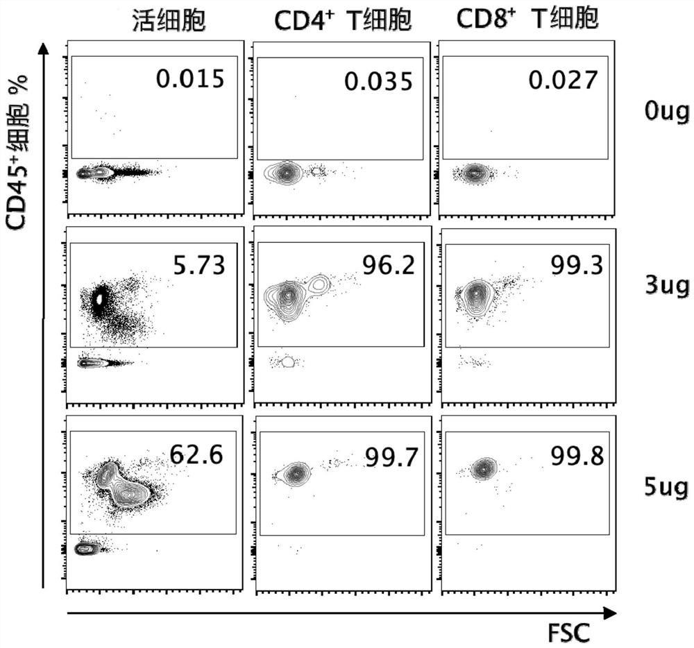
[0042] Embodiment 1 Antibody injection concentration analysis
[0043] 1 material
[0044] 1.1 Experimental animals
[0045] C57BL / 6J mice, 13-16 weeks old, male, SPF grade, were purchased from Beijing Weitong Lihua Experimental Animal Technology Co., Ltd., and raised in the IVC system of the Animal Center of Xuzhou Medical University Institute of Blood Diseases (Italy TECNIPLAST company, qualified Certificate number: 13103254).
[0046] 1.2 Main reagents and instruments
[0047] Fluorescence-labeled mouse anti-human monoclonal antibodies CD4-PEcy7 (clone No.RM4-5), CD8-APC (clone No.53-6.7), CD45-BV570 (clone No.30-F11), purchased from Biolegend, USA, CD45-APCcy7 (clone No.30-F11) was purchased from BD Company in the United States, and mouse erythrocyte lysate and staining buffer were prepared by ourselves. The flow cytometry instrument model is FACSCalibur (BD Company, USA), and flowjo v10 software is used for data analysis.
[0048] 2 methods
[0049] 2.1 Intravenous ...
Embodiment 2
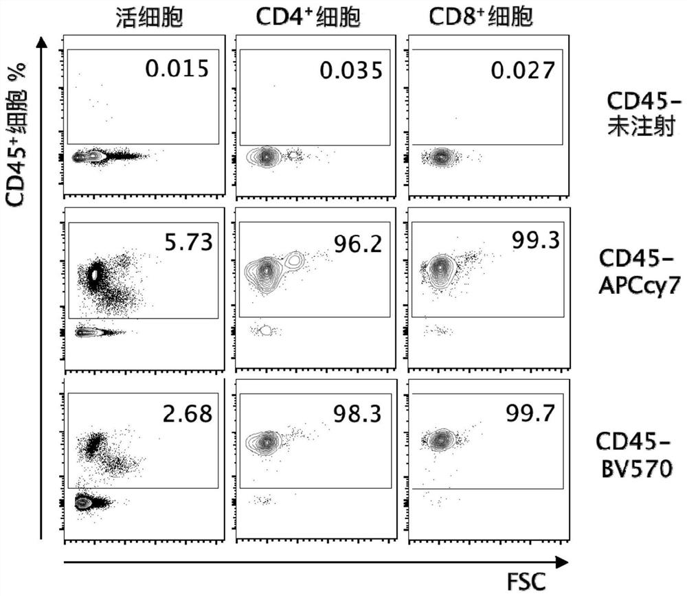
[0053] Example 2 Detection of different fluorescently labeled antibodies
[0054] In order to detect whether antibodies from different markers and manufacturers can achieve similar in vivo staining effects, anti mCD45-APCcy7 and anti mCD45-BV570 antibodies with the same clone number (clone No.30-F11) were used for detection. There was no significant difference in the percentage of CD45+ cells in live cells after injecting 3ug / mouse of anti-mCD45-APCcy7 and anti-mCD45-BV570 antibodies respectively. CD4 stained with two different antibodies + T cells and CD8 + 96% of T cells mean CD45 + cells, the non-staining effect was comparable between the two ( figure 2 ). The above results show that even if the CD45 antibodies with the same clone number are labeled with different fluorescence, it does not affect the ability to quickly bind to the antigen in vivo, and the antibody's ability to recognize intravascular lymphocytes is not affected.
Embodiment 3
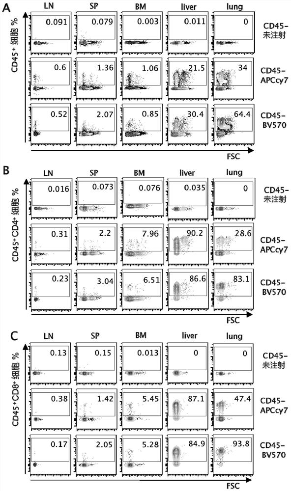
[0055]Embodiment 3 Lymphocyte labeling situation inside and outside blood vessels in lymphoid organs and non-lymphoid organs
[0056] 1 method
[0057] 1.1 Preparation of mononuclear cells
[0058] Peripheral blood mononuclear cells: prepare EDTA anticoagulant EP tubes, add 50ul 0.5M EDTA to 1.5ml EP tubes, remove the mouse eyeballs and take blood and drop them into EP tubes, shake and bounce immediately to prevent coagulation; micropipette draws 200ul peripheral blood mononuclear cells Transfer the blood to a 15ml centrifuge tube, add 5ml mouse erythrocyte lysate to lyse at room temperature for 6 minutes, add 10ml PBS to the centrifuge tube to stop the lysis, centrifuge at 1100rpm, 8min, 4°C, discard the supernatant, add 1ml PBS to the residual solution and place on ice.
[0059] Preparation of LN and SP mononuclear cells: add PBS to the plate without passing through the tissue in the sieve, grind the lymph node and spleen with the needle core of the syringe until only conne...
PUM
 Abstract
Description
Claims
Application Information
 - R&D
- Intellectual Property
- Life Sciences
- Materials
- Tech Scout
- Unparalleled Data Quality
- Higher Quality Content
- 60% Fewer Hallucinations
Browse by: Latest US Patents, China's latest patents, Technical Efficacy Thesaurus, Application Domain, Technology Topic, Popular Technical Reports.
© 2025 PatSnap. All rights reserved.Legal|Privacy policy|Modern Slavery Act Transparency Statement|Sitemap|About US| Contact US: help@patsnap.com