
Establishment method and application of tumor cell three-dimensional model

A tumor cell and three-dimensional model technology is applied in the field of three-dimensional tumor cell culture, which can solve the problems of complex protein fusion and modification methods, unsuitable three-dimensional tumor cell culture, and cell damage.

Pending Publication Date: 2020-08-28
DONGHUA UNIV
View PDF4 Cites 1 Cited by
  • Summary
  • Abstract
  • Description
  • Claims
  • Application Information

AI Technical Summary

Problems solved by technology

[0004] Patent CN201910789298.1 introduces a degradable microsphere with a microtopological structure on the surface. The elliptical microsphere with a concave or wrinkled structure on the surface is prepared by an emulsification method. The particle size of the microsphere described in this patent is 1 to 600 microns. It has good biocompatibility and cells can adhere and grow on the surface of the microspheres, but the degradation time of the microspheres is too long (the degradation residual rate of 3 months is still 85% to 98%), which is not suitable for tumor cell proliferation. Three-dimensional culture; patent CN201510072086.3 introduces a three-dimensional composite microsphere, using natural or chemically synthesized polymers to prepare submicron microspheres, and modifying the microspheres with human E-cadherin to prepare surface-modified E-calcium Adhesin-mediated biomaterial microspheres, the microspheres described in this patent can promote the co-cultivation of microspheres and cells through cadherin and adherens junction signal molecules to form microspheres / stem cell composite microspheres for the proliferation of stem cells in a three-dimensional state , differentiation research, but the protein fusion and modification methods used in the preparation of microspheres by this method are relatively complicated, and E-cadherin only exists on the surface of the microspheres and cannot interact with the cells inside the microspheres. Methods such as hanging drop or centrifugation, these processes will cause certain damage to the cells; patent CN201910133058.6 introduces a cell-carrying alginate gel microsphere, the matrix material is a natural polymer material alginate solution and a divalent metal Ions undergo chelation reaction to prepare hydrogel, in which the gel system is spherical rubber beads with a particle size of 100-2000 microns. The microspheres described in this patent can make cells grow in a three-dimensional way, and are more economical than animal models. However, the microsphere lacks biological factors to maintain cell activity, and has disadvantages such as unstable cell activity and poor clustering of tumor cells when embedding cells.

Method used


Image

  • Establishment method and application of tumor cell three-dimensional model
  • Establishment method and application of tumor cell three-dimensional model

Examples


Embodiment 1

[0034] A method for establishing a three-dimensional model of tumor cells, the steps are as follows:

[0035] (1) Prepare platelet-rich fibrin (PRF) powder and store aseptically;

[0036] The preparation process of platelet-rich fibrin powder is as follows: Use disposable sterile blood collection tube to draw 10ml of venous blood from rabbit ears, place it in a sterile centrifuge tube, and quickly centrifuge at 2500r / min for 10min, and let it stand for 1h before taking coagulation. The yellow gel layer in the middle of the material is made into granules and stored at 4℃ for later use;

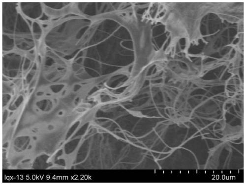
[0037] The average diameter of the platelet-rich fibrin powder is 200 microns, and the scanning electron microscope structure is like figure 1 Shown

[0038] (2) Prepare platelet-rich fibrin powder-collagen composite porous microspheres and store them aseptically;

[0039] (2.1) Prepare a collagen solution with a concentration of 1.2wt%;

[0040] (2.2) Add the platelet-rich fibrin powder to the collagen ...

Embodiment 2

[0052] A method for establishing a three-dimensional model of tumor cells, the steps are as follows:

[0053] (1) Prepare platelet-rich fibrin powder and store aseptically;

[0054] The preparation process of platelet-rich fibrin powder is as follows: Take appropriate amount of venous blood as needed, place it in a sterile centrifuge tube, and quickly centrifuge at 3000r / min for 15min. After standing for 1h, take the yellow gel layer in the middle of the coagulum to make particles ,Store at 4℃ for later use;

[0055] The average diameter of platelet-rich fibrin powder is 250 microns;

[0056] (2) Prepare platelet-rich fibrin powder-collagen composite porous microspheres and store them aseptically;

[0057] Using microfluidic method, the collagen solution dispersed with platelet-rich fibrin powder is made into platelet-rich fibrin powder-collagen composite porous microspheres with an average diameter of 480 microns, an average pore size of 11 microns, and a porosity of 48%;

[0058] When...


PUM

PropertyMeasurementUnit
The average diameteraaaaaaaaaa
Average pore sizeaaaaaaaaaa

Abstract

The invention relates to an establishment method and application of a tumor cell three-dimensional model, and the establishment method comprises the following steps: (1) preparing platelet-rich fibrinpowder, and carrying out aseptic preservation; (2) preparing platelet-rich fibrin powder-bioactive material composite porous microspheres, and carrying out aseptic preservation; and (3) co-culturingthe tumor single cells and the composite porous microspheres to obtain a tumor cell three-dimensional model, wherein the degradation starting time of the bioactive material is shorter than the co-culture time. The application is as follows: the tumor cell three-dimensional model is used as an anti-tumor drug screening model or used for establishing a mouse tumor pathological model. The establishment method disclosed by the invention is simple, has excellent component simulation on tumor microenvironments such as tumor extracellular matrix and the like, can be used for rapidly evaluating the drug effect of the anti-tumor drug in a high-throughput manner, greatly improves the preclinical prediction efficiency and is easy to popularize.

Description

Technical field [0001] The invention belongs to the field of three-dimensional culture of tumor cells, and specifically relates to a method and application for establishing a three-dimensional model of tumor cells. Background technique [0002] Anti-tumor drug resistance in patients with advanced cancer has led to a high cancer mortality rate. Therefore, the development of new anti-tumor drugs is essential to improve patient survival. The rapid and effective screening of new anti-tumor drugs relies on more predictive preclinical research models, and follow-up animal experiments and human clinical trials are performed on the drugs obtained through screening to improve the efficiency of research and clinical transformation. The tumor microenvironment plays a leading role in regulating tumor occurrence, progression, and metastasis. The highly dynamic interaction between tumor cells and the surrounding environment indicates that studying tumors in biomaterial scaffolds may be more s...

Claims


Application Information

Patent Timeline
IPC IPC(8): G06T17/00G16H50/50
CPCG06T17/00G16H50/50
Inventor 李超婧刘星星胡梦博徐晨阳王富军王璐
Owner DONGHUA UNIV
Learn More