
Amphiphilic DNA nano-micelle and preparation method and application thereof

A nanomicelle, amphiphilic technology, applied in the field of biofluorescence analysis, can solve the problems of affecting the stability of the reaction system, limiting the application value, and unable to directly realize the specificity and quantitative detection of biomolecules. Selective, reproducible effects

Active Publication Date: 2020-08-18
SOUTHWEST UNIVERSITY
View PDF11 Cites 2 Cited by
  • Summary
  • Abstract
  • Description
  • Claims
  • Application Information

AI Technical Summary

Problems solved by technology

However, traditional amphiphilic copolymer micelles release internal molecules through UV or pH regulation. These regulation methods not only cannot directly achieve the specific and quantitative detection of biomolecules, but also may be induced by long-term UV radiation or pH environment. Changes affect the stability of the reaction system, which limits its application value to a certain extent

Method used


Image

  • Amphiphilic DNA nano-micelle and preparation method and application thereof
  • Amphiphilic DNA nano-micelle and preparation method and application thereof
  • Amphiphilic DNA nano-micelle and preparation method and application thereof

Examples


Embodiment 1

[0029] Preparation of Amphiphilic DNA Nanomicelles

[0030] In this example, the target miRNA is miRNA-21

[0031] 1) Design the specific DNA hairpin sequence H1-H5 (SEQ ID NO: 1-5) of the target miRNA-21, and modify the hydrophobic C12 alkane chain (Spacer C12) at the end of the H3 hairpin sequence by specific chemical coupling technology 10 , to obtain amphiphilic DNA copolymer sp-H3; synthesized by trivalent phosphorus chemical technology to obtain freeze-dried powder of H1, H2, H4, H5 and sp-H3.

[0032] 2) The preparation of the amphiphilic DNA copolymer sp-H3 dispersion with hairpin structure, the specific steps are as follows:

[0033] First, the sp-H3 freeze-dried powder was centrifuged at 12000r / min for 10min, and then dissolved in a solution containing Mg 2+ In the TE buffer, make it uniformly dispersed to obtain a sp-H3 solution with a concentration of 5nM. Then put the sp-H3 solution into 100 μL EP tubes, place it in a PCR instrument and incubate at 95°C for 10 ...

Embodiment 2

[0036] Label-free detection of miRNA-21 by amphiphilic DNA nanomicelles

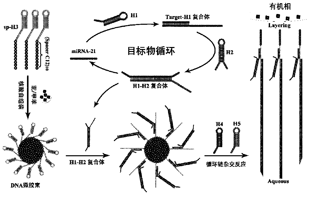
[0037] The principle of label-free detection of miRNA-21 (SEQ ID NO: 6) by DNA nanomicelle is as follows figure 1 As shown, the amphiphilic DNA copolymer sp-H3 is obtained by modifying the hydrophobic C12 alkane chain at the end of the hydrophilic DNA, and the sp-H3 forms a hairpin structure for wrapping the fat-soluble dye perylene. The hydrophilic and hydrophobic interactions between them assemble into stable DNA nanomicelles. In the presence of the target miRNA-21, the catalytic hairpin self-assembly reaction (CHA) between DNA hairpins H1 and H2 is triggered to realize the recycling of trace targets, and at the same time export a large amount of H1-H intermediates; the intermediate The body can open the sp-H3 on the DNA nanomicelle, exposing the cohesive end of the sp-H3. Furthermore, the sticky end of sp-H3 can trigger the hybridization chain reaction (HCR) between H4 and H5, thereby enhancing the ...


PUM


Abstract

The invention belongs to the technical field of bioluminescence analysis, and particularly relates to an amphiphilic DNA nano-micelle and a preparation method and application thereof. The preparationmethod comprises the following steps: 1, designing a specific DNA hairpin sequence H1-5 of a target miRNA, modifying a hydrophobic C12 alkane chain at the tail end of H3 to prepare sp-H3 freeze-driedpowder, preparing a sp-H3 dispersion liquid with a hairpin structure from the obtained sp-H3 freeze-dried powder, finally adding a fluorescent dye into the dispersion liquid, and carrying out ultrasonic treatment to obtain the DNA nano-micelle. The invention also provides an application of the DNA micelle nanometer in label-free detection of target miRNA. The DNA micelle constructed by the invention not only can be quickly assembled into a spherical nano structure, but also has relatively large immobilization capacity, meanwhile, the amphiphilic DNA nano-micelle has the characteristics of goodselectivity, convenience in operation and the like, and has important significance for measuring small biological molecules.

Description

technical field [0001] The invention belongs to the field of bioluminescence analysis, and in particular relates to an amphiphilic DNA nano-micelle and its preparation method and application. Background technique [0002] As a highly controllable emerging material in the field of nanotechnology, DNA nanostructures have attracted much attention, and have great development potential in drug delivery, disease diagnosis, and early detection of tumor markers. In recent years, various DNA nanostructures, such as DNA tetrahedron, DNA cube, etc., have been designed as nanocages for immobilizing small molecules for high-sensitivity analysis and detection. However, the DNA sequence used to assemble nanostructures usually takes a lot of time to carefully design. At the same time, the conditions for assembling nanostructures from long DNA chains are relatively harsh, and the assembly time is long and the assembly efficiency is low, which limits the DNA-based assembly. Wide range of app...

Claims


Application Information

Patent Timeline
IPC IPC(8): C12Q1/6811C12Q1/6813B82Y5/00
CPCC12Q1/6811C12Q1/6813B82Y5/00C12Q2525/113C12Q2525/301C12Q2563/107
Inventor 梁文斌彭锌卓颖柴雅琴袁若
Owner SOUTHWEST UNIVERSITY
Learn More