
Lung cell pathology rapid on-site evaluation system and method and computer readable storage medium

A cytopathology and evaluation system technology, applied in computer parts, calculation, pathological reference, etc., can solve the problems of time-consuming, wasting time, prolonging the ROSE time, etc., and achieve the effect of improving the efficiency of diagnosis

Pending Publication Date: 2020-08-04
SHANGHAI XINGMAI INFORMATION TECH CO LTD
View PDF5 Cites 9 Cited by
  • Summary
  • Abstract
  • Description
  • Claims
  • Application Information

AI Technical Summary

Problems solved by technology

However, there is such a technical defect in the prior art that the physician who performs ROSE on bronchoscopy biopsy prints and fine-needle aspiration smears must be a pathologist, and it is difficult for respiratory physicians to complete ROSE. The needle puncture smear is sent to the pathology department. This process will prolong the time of ROSE. While the patient is on the operating table, the respiratory doctor is often performing the operation. There is an urgent need for real-time detection and evaluation of the structure. Time on the operating table is very precious, and the current microscope scanner will generate a complete microscopic image after scanning the entire bronchoscope biopsy print or fine needle aspiration smear, and cannot Scanning and processing evaluation pictures, which prolongs the time of evaluation and processing, and brings difficulties to the rapid diagnosis of the disease
All in all, the current problem is that it needs to be sent to the pathology department for manual inspection, which is time-consuming, and the second is that it is time-consuming to start the inspection after the large-scale image is collected.

Method used


Image

  • Lung cell pathology rapid on-site evaluation system and method and computer readable storage medium
  • Lung cell pathology rapid on-site evaluation system and method and computer readable storage medium
  • Lung cell pathology rapid on-site evaluation system and method and computer readable storage medium

Examples


Embodiment Construction

[0035] In order to better illustrate the invention purpose of the present invention, the implementation of the technical solution and the advantages of the present invention compared with the prior art, the following will specifically combine the illustrated drawings and examples of different embodiments in an exemplary manner , the present invention is further elaborated and illustrated. It should be understood that the specific embodiments described or exemplified in this section are only used to explain or facilitate the understanding of the overall inventive concept of the present invention, but should not be construed as limiting the protection scope of the claims of the present invention. All the inventive concept and core of the present invention fall within the protection scope of the present invention, especially the equivalent replacement or specific deformation based on the inventive concept or theme of the present invention will all fall within the protection scope ...


PUM


Abstract

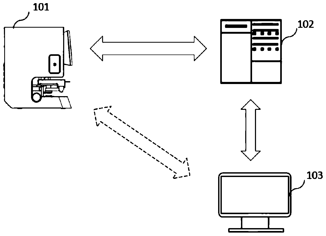
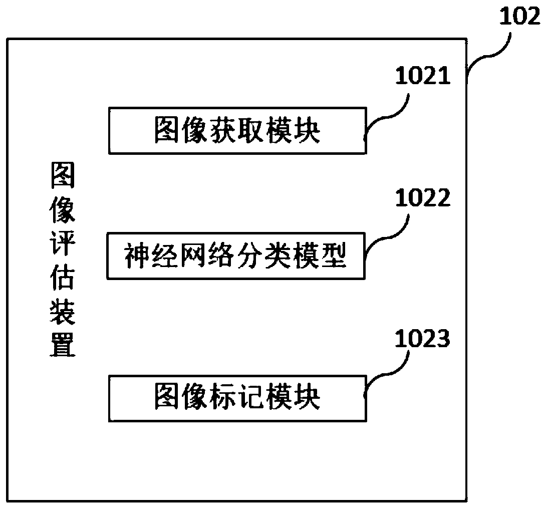
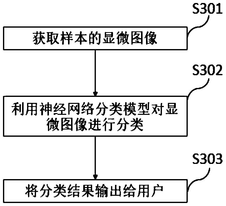
The invention provides a lung cell pathology rapid on-site evaluation system and method and a computer readable storage medium, which are used for rapidly evaluating a cell sample on an operation site. The lung cell pathology rapid on-site evaluation system comprises a microscopic image acquisition device which comprises an objective table used for bearing a lung cell sample and a camera used forshooting the cell sample to obtain a microscopic image of the sample; an image evaluation device with a neural network classification model used for classifying the microscopic images, wherein the obtained classification result is one of lung squamous cell carcinoma, lung adenocarcinoma, small cell lung cancer, unclear non-small cell lung cancer, other malignant lesions, no obvious abnormality, granuloma and inflammation; and an output device connected to the image evaluation device and used for outputting the classification result to a user. According to the invention, the neural network classification model is used for evaluating the microscopic image acquired by the microscopic image acquisition device, and an evaluation result is obtained on an operation site, so that the problems thatthe current cell pathological diagnosis is complex and time-consuming, and the pathological diagnosis result cannot be obtained immediately are solved, and the diagnosis efficiency is effectively improved.

Description

technical field [0001] The invention relates to an image processing technology, in particular to a rapid on-site assessment and evaluation technology of lung cytopathology, and belongs to the technical field of microscopic pathological auxiliary diagnosis. Background technique [0002] At present, the method of auxiliary diagnosis of the lung is usually a needle biopsy guided by computed tomography (CT), which is a relatively common method for diagnosing the lung. The present invention is mainly aimed at collecting specimen cells at bronchi or lung lesion positions through fiberoptic bronchoscope lung biopsy to further evaluate and process them, so as to assist in the diagnosis of diseases. Currently, rapid on-site evaluation of specimens is commonly used in this field, that is, ROSE (Rapid on-site evaluation), which refers to rapid on-site inspection of specimen cells by cytopathologists and evaluation of the quality of fine-needle aspiration smears and biopsy tissue prints...

Claims


Application Information

Patent Timeline
IPC IPC(8): G16H70/60G06K9/00G06K9/62G06N3/04G06N3/08
CPCG16H70/60G06N3/08G06V20/698G06N3/045G06F18/24
Inventor 叶德贤房劬姜辰希
Owner SHANGHAI XINGMAI INFORMATION TECH CO LTD
Learn More