
Fundus image quality evaluation method based on blood vessel segmentation and background separation

A fundus image and quality evaluation technology, which is applied in the field of image quality evaluation based on convolutional neural network, can solve problems such as delaying treatment timing, misdiagnosis is not easy to be found, and image display is insufficient for medical diagnosis, so as to improve expression ability and weaken sensitivity Effect

Active Publication Date: 2020-08-04
ZHEJIANG UNIV OF TECH
View PDF5 Cites 8 Cited by
  • Summary
  • Abstract
  • Description
  • Claims
  • Application Information

AI Technical Summary

Problems solved by technology

A study involving an extensive database of retinal images reveals that more than 25 percent of images appear to be of insufficient quality for proper medical diagnosis
In addition to the financial investment required to reacquire poor quality photographs, it is inconvenient for the patient to return to the medical center for repeat fundus photographs
What's more serious is that misdiagnosis caused by poor image quality is not easy to be found, delaying the timing of treatment

Method used


Image

  • Fundus image quality evaluation method based on blood vessel segmentation and background separation
  • Fundus image quality evaluation method based on blood vessel segmentation and background separation

Examples


Embodiment Construction

[0015] The present invention will be further described below in combination with schematic diagrams.

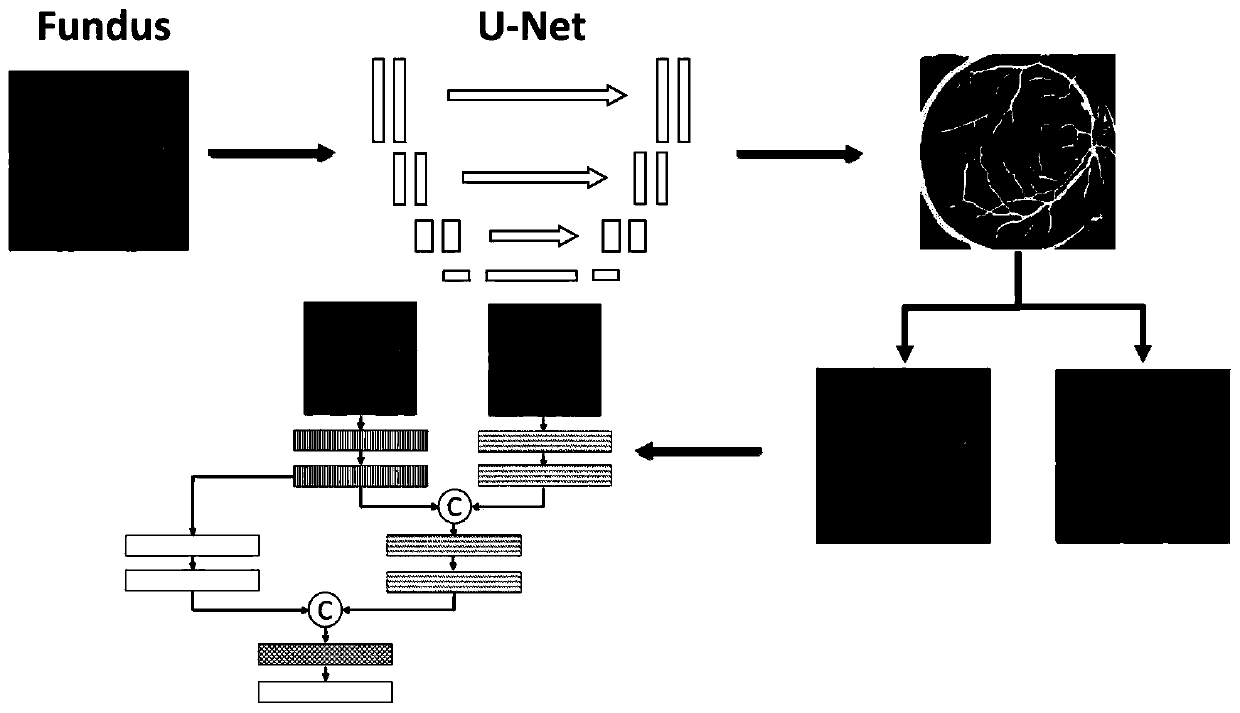
[0016] refer to figure 1 and figure 2 , a background separation fundus image quality assessment method based on blood vessel segmentation guidance, comprising the following steps:

[0017] 1) First, blood vessel segmentation is performed on the input image through the U-Net model pre-trained on the DRIVE public fundus image dataset;

[0018] 2) Multiply the blood vessel feature map obtained in step 1) with the original image element by element to obtain an image containing only blood vessel and background information;

[0019] 3) Use the extracted feature images to input the network branches respectively for training to obtain model parameters;

[0020] 4) The trained convolutional neural network model is used to evaluate the quality of the test pictures.

[0021] Further, the network structure implemented in step 3) includes two parts: a dual-branch feature extraction p...


PUM


Abstract

The invention discloses a fundus image quality evaluation method based on blood vessel segmentation and background separation, and the method comprises the following steps: 1), carrying out the bloodvessel segmentation of an input image through a pre-trained U-Net model on a DRIVE public fundus image data set; 2) multiplying the blood vessel feature map obtained in the step 1) by an original image element by element to obtain an image only containing blood vessels and background information; 3) respectively inputting the extracted feature images into convolutional neural network branches fortraining to obtain model parameters; and 4) performing quality evaluation on the test picture by using the trained convolutional neural network model. According to the invention, higher evaluation accuracy is realized, the reexamination rate of doctors is reduced, and possible treatment opportunity delay caused by repeated examination is avoided. The model provided by the invention has universality and can be embedded into various advanced convolutional neural network structures, the network performance is improved, and meanwhile, a method for fusing vascular prior knowledge and neural networkend-to-end feature extraction is provided.

Description

technical field [0001] The invention relates to the fields of medical image processing and computer vision, in particular to an image quality evaluation method based on a convolutional neural network. Background technique [0002] The fundus image is captured by a special fundus camera, which contains important physiological structures such as the optic disc, macula, and blood vessels in the retina, and is an important type of image in medical imaging. In the normal fundus image, the optic disc appears as a nearly circular bright color area, which has the strongest contrast with the background area, and is the starting area of ​​the optic nerve and blood vessels; because the macula is rich in lutein, it is in the normal fundus image It is manifested as a dark area, and the dark area has no blood vessel structure, and there is an inwardly sunken area called the fovea in the center of the macula; the blood vessels start from the optic disc area and extend to the entire interio...

Claims


Application Information

Patent Timeline
Patent Type & Authority Applications(China)
IPC IPC(8): G06T7/00G06T7/11G06T7/194G06N3/04G06N3/08
CPCG06T7/0012G06T7/11G06T7/194G06N3/08G06T2207/10004G06T2207/20081G06T2207/20084G06T2207/30041G06T2207/30168G06N3/045
Inventor 刘义鹏吕雅俊王海霞蒋莉陈朋梁荣华
Owner ZHEJIANG UNIV OF TECH
Learn More