Method for determining chromosome structure variation signal intensity and insert fragment length distribution characteristics of sample, and application thereof
A technology of inserting fragments and structural variation, applied in the field of bioinformatics, can solve problems such as chromosomal rearrangement and miscarriage
- Summary
- Abstract
- Description
- Claims
- Application Information
AI Technical Summary
Problems solved by technology
Method used
Image

Examples
Embodiment 1
[0077] Example 1 Sample plasma separation, library preparation, sequencing on the machine;
[0078] 1. Plasma Separation
[0079] a) Prepare the instruments, reagents, and consumables required for the experiment, and the high-speed refrigerated centrifuge should be pre-cooled to 4°C in advance.
[0080] b) If the peripheral blood sample is collected with an EDTA anticoagulant tube, put it in a 4°C refrigerator immediately after drawing the blood, and conduct plasma separation within 2 hours. If the peripheral blood sample is collected with free nucleic acid storage tubes such as streck tubes, it can be placed at room temperature, and plasma separation can be performed within the time specified in the blood collection tube instructions.
[0081] c) Record the sample information, balance the blood collection tube, replace the high-speed refrigerated centrifuge with a horizontal rotor, and set parameters: temperature 4°C, centrifugal force 1600g, time 10min. Place the blood col...
Embodiment 2
[0190] 1. According to the method of Example 1, complete the library sequencing of the samples, obtain off-machine data, filter out low-quality reads, and use the comparison software (bwa) to compare these sequencing reads to the human reference genome ( hg19).
[0191] Second, compare and compare the bam files for statistics:
[0192] 1. Filter reads and duplicate reads with only one end alignment (duplicate reads marked with samtools or picard)
[0193] 2. Statistical low-quality alignment rate: low-quality alignment refers to the alignment reads whose alignment quality value of reads at any end is less than 30. Add up all these reads and divide by the total number of reads, which is the low-quality alignment Rate;
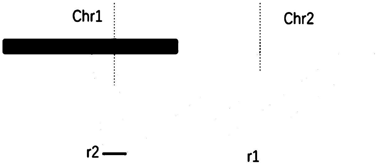
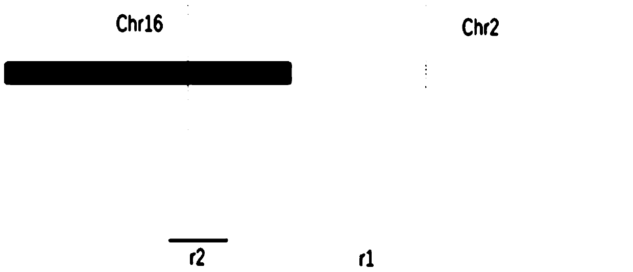
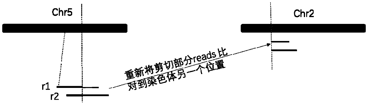
[0194] 3. For abnormal alignments with high alignment quality values (greater than 30), count the signal strength of reads supporting structural variation: including 1) as follows Figure 4 As shown, the statistics of the number of reads (reads) in the thre...
Embodiment 3
[0243] Based on Example 2, it is obtained: (1) the signal intensity supporting the variation of chromosome structure; (2) the ratio of mitochondrial content of the sample; (3) the ratio of inserts in the entire sample between 180-220 and 250-300, and those less than 150 bp The "peak-to-valley spacing" between the peaks and troughs. (3) The ratio of the number of short fragments to long fragments in each 5M interval, after dimensionality reduction by principal component analysis, take the value of the first 10 principal components.
[0244] Input these statistical values of samples as feature vectors, use machine learning methods (such as: SVM, Lasso, GBM), and based on the above nearly 400 cancer and normal samples, use 10-fold cross-validation to test the effect of tumor prediction. The samples were divided into 10 points on average, and 9 of them were used as the training set in order to establish a tumor prediction model. The remaining one is used as a training set to me...
PUM
 Abstract
Description
Claims
Application Information
 - R&D
- Intellectual Property
- Life Sciences
- Materials
- Tech Scout
- Unparalleled Data Quality
- Higher Quality Content
- 60% Fewer Hallucinations
Browse by: Latest US Patents, China's latest patents, Technical Efficacy Thesaurus, Application Domain, Technology Topic, Popular Technical Reports.
© 2025 PatSnap. All rights reserved.Legal|Privacy policy|Modern Slavery Act Transparency Statement|Sitemap|About US| Contact US: help@patsnap.com