Eureka AIR delivers breakthrough ideas for toughest innovation challenges, trusted by R&D personnel around the world.

A Multi-organ Segmentation Method of Thoracic Cavity Based on Cascaded Residual Fully Convolutional Network

A fully convolutional network and multi-organ technology, applied in the field of thoracic multi-organ segmentation based on the cascaded residual full convolutional network, can solve the problem of being easily limited by hardware capabilities, excessive 3D network calculations, and inability to learn spatial information and other problems to achieve accurate segmentation, stable training process, and good training effect

Active Publication Date: 2022-04-29
ZHEJIANG UNIV
View PDF8 Cites 0 Cited by
  • Summary
  • Abstract
  • Description
  • Claims
  • Application Information

AI Technical Summary

Problems solved by technology

One is a method based on a 2D model. This method cuts a 3D CT image into a 2D image, and uses a 2D segmentation network, such as u-net, to perform segmentation, and superimposes the obtained multiple 2D segmentation results to obtain a 3D image. Segmentation results. The advantage of this method is that it can accurately segment each layer of images and learn more detailed information. However, it cannot learn spatial information of another dimension, and the spatial continuity is poor.
Another method is the method based on the 3d model. This method directly inputs the CT image into the 3d segmentation network, such as v-net, 3d u-net, etc., and directly outputs the 3d segmentation result. The advantage of this method is that it can take into account The association between layers, however, 3D network calculation is too large, easily limited by hardware capabilities

Method used


Image

  • A Multi-organ Segmentation Method of Thoracic Cavity Based on Cascaded Residual Fully Convolutional Network
  • A Multi-organ Segmentation Method of Thoracic Cavity Based on Cascaded Residual Fully Convolutional Network
  • A Multi-organ Segmentation Method of Thoracic Cavity Based on Cascaded Residual Fully Convolutional Network

Examples


Embodiment Construction

[0035] The present invention will be further described in detail below with reference to the accompanying drawings and embodiments. It should be noted that the following embodiments are intended to facilitate the understanding of the present invention, but do not limit it in any way.

[0036] In the specific embodiment of the present invention, the multi-organ segmentation of thoracic CT is taken as an example to illustrate, but the present invention is not limited thereto. Non-essential improvements and adjustments made by those skilled in the art under the core guiding ideology of the present invention still belong to protection scope of the present invention.

[0037] Step 1: Data preprocessing in the rough segmentation stage

[0038]This article uses chest CT images. In order to remove excessive impurities and improve the contrast between organs and the background, in the rough segmentation stage, we cut off the range of CT values, set the window level to -500, and the win...


PUM


Abstract

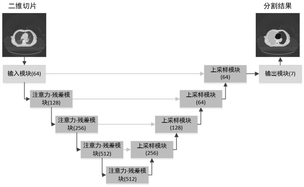
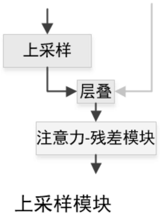
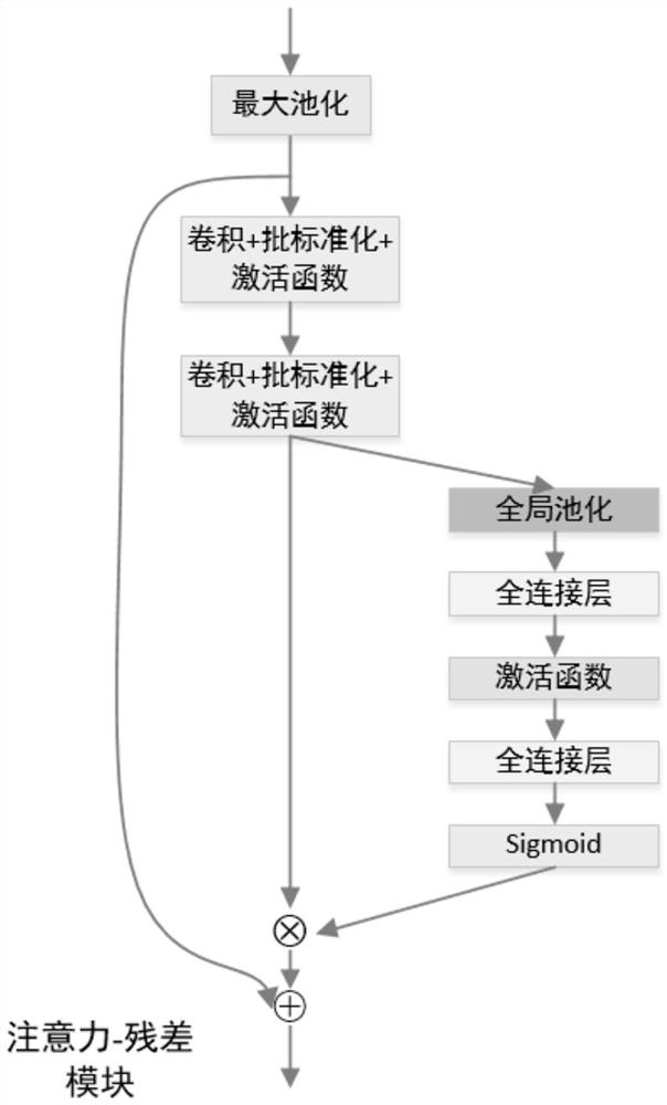
The invention discloses a multi-organ segmentation method of the thoracic cavity based on a cascaded residual full convolution network. First, a rough segmentation model is used to perform a rough segmentation of each organ, and the function is to locate the range of each organ, and then, use the Separate the fine segmentation model, perform fine segmentation on the area of ​​coarse segmentation and positioning, and obtain the fine segmentation results of each organ, and finally combine the results to obtain the final multi-organ segmentation result, which retains more details. The present invention improves on the basis of u-net, introduces the residual connection and the attention mechanism of the feature dimension into the down-sampling module in u-net, makes the network easier to train, and has the ability of automatic feature selection. In addition, we introduced a cascading strategy and used a staged segmentation network to achieve fast and accurate segmentation of multiple organs in chest CT.

Description

technical field [0001] The invention belongs to the field of medical CT image processing, and in particular relates to a multi-organ segmentation method of the thoracic cavity based on a cascaded residual full convolution network. Background technique [0002] Radiation therapy is a common option for treating lung and esophageal cancer. During radiation therapy planning, physicians will manually delineate the target tumor and nearby organs (known as organs at risk OARs), which is often time-consuming and plagued by large subjective variability. To make matters worse, certain organs, such as the esophagus and trachea, are difficult to delineate due to large variations in position and contour, small size, and low contrast in CT scans. [0003] CT is the most commonly used detection method in disease diagnosis. It uses X-ray beams to scan a layer of a certain thickness of the human body, and the detector receives the X-rays that pass through this layer, converts them into visi...

Claims


Application Information

Patent Timeline
Patent Type & Authority Patents(China)
IPC IPC(8): G06T7/11G06T3/40G06N3/08G06N3/04
CPCG06T7/11G06T3/4007G06N3/08G06T2207/10081G06T2207/20132G06N3/045
Inventor 吴健雷璧闻应豪超余柏翰
Owner ZHEJIANG UNIV
Why Eureka
Eureka Blog
Learn More