
Method for detecting and positioning of cancer regions with small samples or unbalanced samples

A technology of area detection and small samples, which is applied in the direction of neural learning methods, instruments, biological neural network models, etc., can solve problems such as gradient disappearance, over-fitting, and too many parameters, so as to improve the accuracy of discrimination and solve the problem of low accuracy problems and improve diagnostic efficiency

Inactive Publication Date: 2019-12-31
SICHUAN CHANGHONG ELECTRIC CO LTD
View PDF12 Cites 17 Cited by
  • Summary
  • Abstract
  • Description
  • Claims
  • Application Information

AI Technical Summary

Problems solved by technology

The iterative training model of the invention uses the convolutional neural network VGG+support vector machine for the final multi-classification problem. Although increasing the depth of the neural network will improve the performance of the network, blindly increasing the depth of the network will bring two disadvantages: the parameter Too many, in the face of the problem addressed by the present invention: when the data set is small, it is easy to cause overfitting, and the problem of gradient disappearance is easy to occur if the number of layers is too deep. In addition, this method is based on video content rather than large digital image data Therefore, it focuses on the targeted collection of data, the use of cascaded character representations of different body parts and different granularities, and the targeted training of behavioral classifiers, which is not suitable for cancer cell identification and detection
A coarse-to-fine analysis idea based on pathological images also proposed by Chekkoury et al. ("Automated malignancy detection in breast histopathological images", Proc.SPIE Med.Imag., vol.8315, pp.831515-1-831515-13 , 2012.), but the disadvantage of this idea is that it requires human-labeled features, which cannot well cover other tumor types

Method used


Image

  • Method for detecting and positioning of cancer regions with small samples or unbalanced samples
  • Method for detecting and positioning of cancer regions with small samples or unbalanced samples
  • Method for detecting and positioning of cancer regions with small samples or unbalanced samples

Examples


Embodiment Construction

[0040] In order to make the purpose, technical solution and advantages of the present invention clearer, the technical solution of the present invention will be described in detail below. Apparently, the described embodiments are only some of the embodiments of the present invention, but not all of them. Based on the embodiments of the present invention, all other implementations obtained by persons of ordinary skill in the art without making creative efforts fall within the protection scope of the present invention.

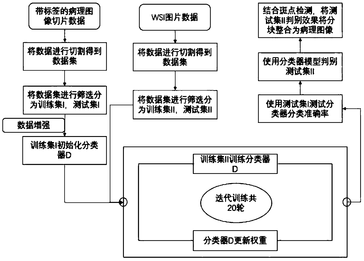
[0041] In either embodiment, if figure 1 As shown, the method for detecting and locating cancer regions with small samples or unbalanced samples of the present invention, the specific implementation method includes the following steps:

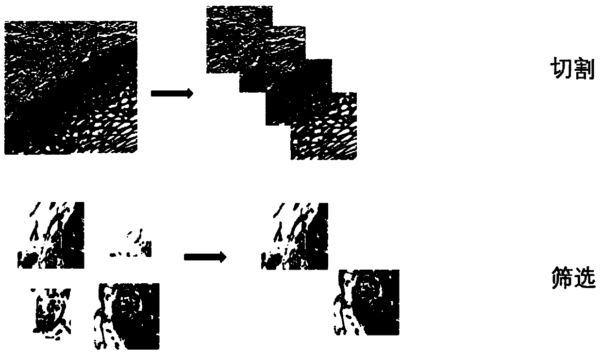
[0042] Step 1: Data preprocessing. The present invention uses a total of two types of image data, one is labeled cervical cancer cell histopathological image slice data, using the training set to initialize the weak classifier...


PUM


Abstract

The invention discloses a method for detecting and positioning of cancer regions with small samples or unbalanced samples. By combining the characteristics of the histopathological image containing cancer cells, the method enhances the data set by using a method of adding noise, rotating, increasing or reducing brightness, expands the data set, balances the specific gravity of the label type of the training set, and improves the training effect of the classifier. The method is characterized in that an Inception V2 network is used as a basis; aiming at the conditions of few samples and unbalanced samples, iterative training is carried out in combination with a small number of calibrated cancerous tissue image blocks and WSI pathological images; the method is advantaged in that WSI image-level pathological image cancer area detection and positioning can be accomplished with high accuracy; problems of over-fitting, local optimization and gradient disappearance of the over-deep neural network easily caused by too few labeled training samples are solved; and accuracy of the training result and usability of the network under the same level are improved.

Description

technical field [0001] The invention relates to the technical field of artificial intelligence deep learning, in particular to a method for detecting and locating cancer regions with small or unbalanced samples. Background technique [0002] Physicians and pathologists usually combine different digital image data (such as the WSI pathological image used in the present invention) to discuss treatment options when studying different cancer diseases of patients. In the process of analyzing pathological images and making diagnostic decisions by human pathologists, pathologists often need to complete at least dozens or even hundreds of high-power field of view case images classification and statistical work. The entire judgment process is time-consuming and labor-intensive, which is not conducive to patients. In addition, pathologists usually divide the entire WSI into smaller image blocks for more careful labeling and processing. In fact, manual processing is slow at any time an...

Claims


Application Information

Patent Timeline
Patent Type & Authority Applications(China)
IPC IPC(8): G06K9/62G06N3/04G06N3/08
CPCG06N3/08G06V2201/03G06N3/048G06N3/045G06F18/217G06F18/24G06F18/214
Inventor 杨懿龄
Owner SICHUAN CHANGHONG ELECTRIC CO LTD
Learn More