
Method for constructing 3D printed individual orthopeadic implant and enhancing biomechanics of 3D printed individual orthopeadic implant

A 3D printing and biomechanics technology, applied in medical science, computer-aided surgery, internal fixator, etc., can solve problems such as difficult to adapt to high-intensity long-term use, heavy interactive workload, and collapse of diseased bone defects, etc. and automatic optimization, good biomechanical properties, and strong interdisciplinary effects

Inactive Publication Date: 2019-12-03
CHANGZHOU NO 2 PEOPLES HOSPITAL
View PDF10 Cites 14 Cited by
  • Summary
  • Abstract
  • Description
  • Claims
  • Application Information

AI Technical Summary

Problems solved by technology

3D printing needs to print 3D model slices layer by layer. Traditional slicing with equal layer thickness will cause a contradiction between model accuracy and printing time: the layer thickness is small, the model accuracy is guaranteed, but the printing time is long; on the contrary, the printing time is shortened, but the Easy to lead to large model step error
[0020] (1) The 3D bone model reconstruction based on CT images still has modeling difficulties and a large amount of interaction
[0021] (2) For the design of personalized orthopedic implants and surgical guides, it is necessary to be proficient in relevant medical and CAD design software, and to have certain surgical experience, which requires high personnel requirements
[0022] (3) The mechanical properties of 3D printed orthopedic implants have low mechanical strength and poor fatigue resistance, making it difficult to adapt to high-intensity long-term use after surgery
[0024] The 3D bone model reconstruction method based on CT images still has problems such as modeling difficulties and heavy interactive workload
The main reason is that the particularity of medical images makes it difficult for the algorithm to automatically process, for example: the gap between bones is too small, resulting in blurred boundaries between adjacent bones; the gray value of cancellous bone is between bone and soft tissue, making it difficult to distinguish; Collapse of the defect in the diseased bone, causing penetration of the bone border
[0025] The 3D printing optimization of bone models and artificial prostheses not only needs to consider their geometric properties, but also needs to consider anatomical structures and biomechanical characteristics, and the corresponding algorithms have not been reported yet.

Method used


Image

  • Method for constructing 3D printed individual orthopeadic implant and enhancing biomechanics of 3D printed individual orthopeadic implant
  • Method for constructing 3D printed individual orthopeadic implant and enhancing biomechanics of 3D printed individual orthopeadic implant
  • Method for constructing 3D printed individual orthopeadic implant and enhancing biomechanics of 3D printed individual orthopeadic implant

Examples


Embodiment 1

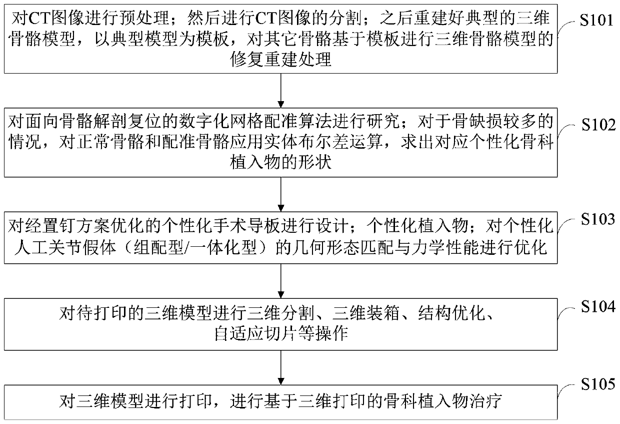
[0113] The 3D printing personalized orthopedic implant construction and biomechanical optimization treatment method provided by the embodiment of the present invention includes the following steps:

[0114] Step 1: Reconstruct and optimize the 3D bone model based on prior knowledge; first, preprocess the CT image; then segment the CT image; then reconstruct a typical 3D bone model, and use the typical model as a template to model other bones based on The template is used for repairing and reconstructing the three-dimensional bone model.

[0115] Step 2: Research on the digital grid registration algorithm for bone anatomical restoration; for the case of many bone defects, apply entity Boolean difference operation to normal bone and registered bone to find the shape of the corresponding personalized orthopedic implant .

[0116] Step 3: Design the personalized surgical guide plate optimized by the nail placement scheme; personalize the implant; optimize the geometric shape matc...

Embodiment 2

[0120] (1) Reconstruction and optimization of 3D bone model based on prior knowledge

[0121] The rapid generation and automatic optimization of 3D bone models are the basis for the application of 3D printing in orthopedic diagnosis and treatment. However, the current quality and automation of 3D bone reconstruction based on CT images is still low. The present invention intends to make full use of prior knowledge of human bone CT images and geometric shapes to improve the accuracy and automation of two-dimensional image segmentation and three-dimensional bone model reconstruction optimization.

[0122] For normal human bones, bones of the same type generally have the same structural features. Therefore, CT images can be preprocessed, a typical three-dimensional bone model can be reconstructed, and the typical model can be used as a template to repair and reconstruct other bones based on the template. In the segmentation of CT images, by establishing a corresponding relations...


PUM


Abstract

The invention belongs to the technical field of image information processing and discloses a method for constructing a 3D printed individual orthopeadic implant and enhancing the biomechanics of the 3D printed individual orthopeadic implant. The method comprises the following steps: firstly, preprocessing a CT image; secondly, cutting the CT image; thirdly, reconstructing a typical three-dimensional skeleton model, taking the typical model as a template, and performing three-dimensional model repairing and reconstructing treatment on other skeletons based on the template; fourthly, studying askeleton anatomical reduction-oriented digital grid registration algorithm; designing an individual surgical template optimized by a screw-setting scheme; optimizing the geometric pattern matching andmechanical performance of an individual artificial joint prosthesis; printing the three-dimensional model, and performing three-dimensional printing-based orthopeadic implant treatment. The method has important theoretical significance and clinical application value in shortening the orthopeadic preoperative planning cycle, improving the implanting efficiency and mechanical performance of individual implants, reducing surgical time and surgical injury and promoting orthopeadic implant industrial development.

Description

technical field [0001] The invention belongs to the technical field of image information processing, and in particular relates to a 3D printing personalized orthopedic internal plant construction and biomechanical optimization processing method. Background technique [0002] At present, the closest existing technology: Before traditional orthopedic surgery, doctors generally understand the condition through X-ray, CT and other imaging methods, and then plan and simulate the operation on two-dimensional drawings or in the mind. This method is difficult to intuitively understand the injured bone area, it is difficult to perform complex surgical simulation, and it has high requirements on the professional level and imagination of doctors. 3D printed mock-ups provide a more realistic environment than digital models. Doctors can make an accurate diagnosis before the operation, formulate a detailed operation plan, evaluate the possible risks during the operation, and practice on ...

Claims


Application Information

Patent Timeline
IPC IPC(8): A61B17/86A61B17/68A61B34/10
CPCA61B17/68A61B17/866A61B2017/00526A61B34/10A61B2034/102A61B2034/105
Inventor 刘宏伟蒋俊锋顾勇张文翁益平徐南伟张云坤魏辉王庆周鑫叠李栋
Owner CHANGZHOU NO 2 PEOPLES HOSPITAL
Learn More