
NMR-optical dual mode imaging biconical DNA nanoprobe and its preparation method and application

A dual-mode imaging and nanoprobe technology, applied in the field of nanomedicine, can solve the problems of increasing toxicity, low sensitivity of nuclear magnetic imaging, and improving contrast agents.

Active Publication Date: 2020-12-01
THE NAT CENT FOR NANOSCI & TECH NCNST OF CHINA
View PDF1 Cites 0 Cited by
  • Summary
  • Abstract
  • Description
  • Claims
  • Application Information

AI Technical Summary

Problems solved by technology

However, due to the non-specificity of gadolinium contrast agent and the low sensitivity of MRI, early diagnosis of tumors is difficult
Increasing the dose of contrast media also increases the risk of gadolinium toxic effects in the body

Method used


Image

  • NMR-optical dual mode imaging biconical DNA nanoprobe and its preparation method and application
  • NMR-optical dual mode imaging biconical DNA nanoprobe and its preparation method and application
  • NMR-optical dual mode imaging biconical DNA nanoprobe and its preparation method and application

Examples


Embodiment 1

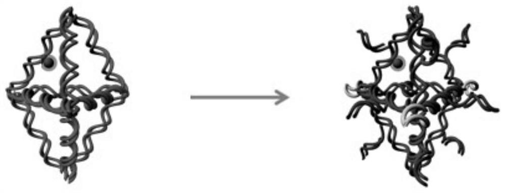
[0101] Example 1 Preparation of NMR-optical dual-modality imaging biconical DNA nanoprobes

[0102] This embodiment adopts the following method to prepare the nuclear magnetic-optical dual-modal imaging biconical DNA nanoprobe, including the following steps:

[0103] 1. Preparation of DNA-DOTA(Gd) MRI contrast agent

[0104] The nuclear magnetic resonance imaging contrast agent is prepared by a two-step organic chemical reaction. The specific steps are as follows: In the first step, the 5'-terminal amino group is modified, and the 3'-terminal Cy3-modified single-stranded DNA is dissolved in deionized water, and the concentration is adjusted to 200 μM; DOTA-NCS is dissolved in 80% DMSO, and the concentration is adjusted to 200 mM; Strand DNA and DOTA-NCS were mixed in 0.1M HEPES (pH=9, containing 300mM MgCl) 2 ) buffer solution, add magnetic stirring, and react in a water bath at 37°C for 24h. This step is the coupling reaction of amino and isothiocyanate. In the second ste...

Embodiment 2

[0126] Example 2 Preparation of NMR-optical dual-modality imaging biconical DNA nanoprobes

[0127] The difference between this example and Example 1 is that the contrast agent for nuclear magnetic resonance imaging selected in the process of synthesizing the double-cone DNA nanoprobe for nuclear magnetic-optical dual-modal imaging is DNA-DTPA (Gd), that is, DTPA and gadolinium are selected. Chelation. Other than that, the selection of other raw materials, the preparation method and the reaction conditions are the same as those in Example 1, so that the nuclear magnetic-optical dual-mode imaging biconical DNA nanoprobe is prepared.

Embodiment 3

[0128] Example 3 NMR-optical dual-modality imaging of live-cell uptake of biconical DNA nanoprobes

[0129] In this example, the following methods were used to investigate the uptake of living cells by the NMR-optical dual-modality imaging biconical DNA nanoprobe, as follows:

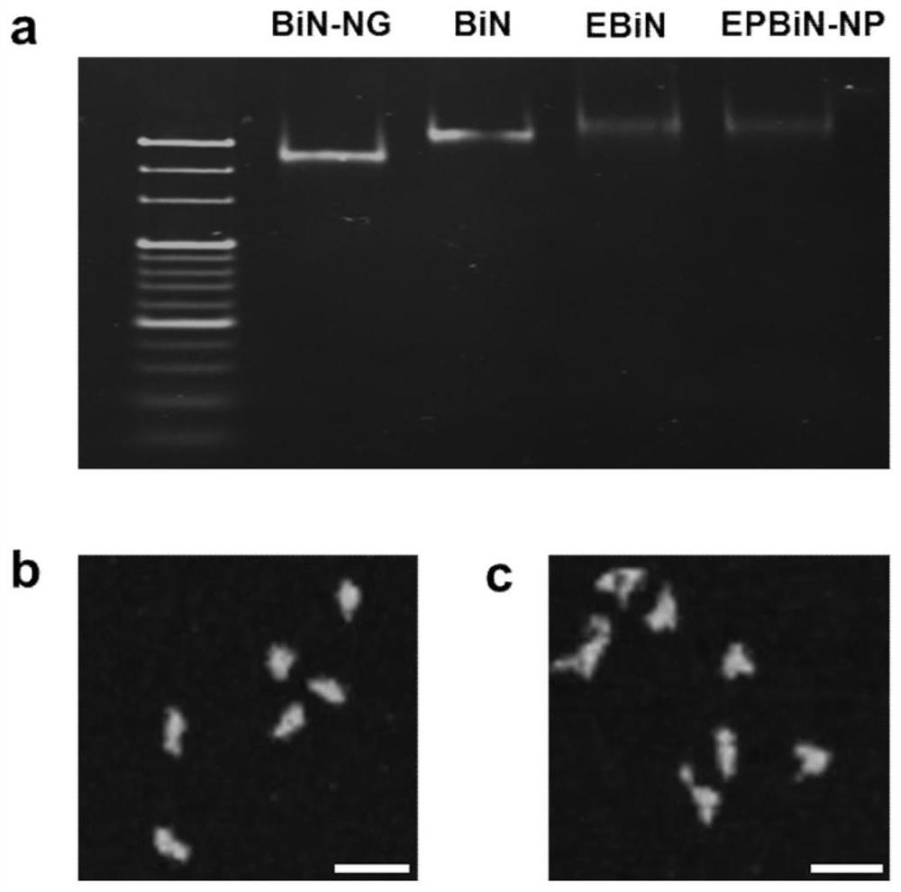
[0130] Due to the excitation wavelength limitation of confocal laser scanning microscopy (CLSM), the modified Dylight800 fluorescent molecule on the DNA probe was replaced with Cy5.5. MDA-MB-231 cells were cultured in 10% fetal bovine serum and 1% double antibody (double antibody is a mixture containing penicillin (10,000IU) and streptomycin (10,000μg / mL) at 100 times the working concentration) complete high-glucose DMEM medium at 37 °C and 5% CO 2 . When the cells grew to about 80% confluence, the cells were digested and re-seeded in 35mm 2 In a confocal dish, the cells were cultured overnight for 12 h. The medium was replaced with a biconical DNA nanostructure (BiN) containing DNA-DOTA(Gd), hybrid...


PUM


Abstract

The invention provides a nuclear magnetism-optical dual-mode imaging biconical DNA nanoprobe, and a preparation method and an application thereof. The dual-mode imaging DNA probe is prepared from a DNA nanostructure, a nucleic acid aptamer, a chemical fluorescent molecule and a nuclear magnetism resonance imaging contrast agent. The DNA probe provided by the invention improves the stability of the nuclear magnetism resonance imaging contrast agent and the chemical fluorescent molecule in body circulation, reduces the potential biological toxicity of gadolinium ions, and improves the targeting ability of tumors by using the nucleic acid adapter, and maintains the T1 relaxation efficiency equivalent to that of a clinical nuclear magnetism resonance contrast agent when the gadolinium content is only 1 / 20 of the clinical dose. With the synergistic effect of magnetic resonance imaging and fluorescence imaging, the DNA probe achieves higher resolution ratio and sensitivity of tumor imaging, and provides powerful technical means for early diagnosis of tumors.

Description

technical field [0001] The invention belongs to the field of nano-medicine, and in particular relates to a nuclear magnetic-optical dual-mode imaging biconical DNA nano-probe and a preparation method and application thereof. Background technique [0002] Magnetic resonance imaging (MRI) is widely used in tumor diagnosis due to its advantages of high imaging resolution, low interference, and imaging in any direction and plane. Magnetic resonance imaging contrast agents refer to preparations that shorten the relaxation times of human tissues such as T1 and T2 to increase the contrast of images, and preparations using paramagnetic or superparamagnetic substances have become the mainstream. The contrast agent is used to amplify all or part of the signal of the desired tissue, or to attenuate the signal of the surrounding tissue, thereby amplifying the light-dark contrast. Gadolinium (Gd) is a rare earth metal element whose atomic orbital contains seven unpaired electrons, makin...

Claims


Application Information

Patent Timeline
Patent Type & Authority Patents(China)
IPC IPC(8): C12N15/11C12N15/115A61K49/00A61K49/10A61K49/12
CPCA61K49/0002A61K49/0032A61K49/0052A61K49/0054A61K49/10A61K49/105A61K49/108A61K49/126C12N15/11C12N15/115C12N2310/16
Inventor 丁宝全王昭然宋琳琳王婷蒋乔
Owner THE NAT CENT FOR NANOSCI & TECH NCNST OF CHINA
Learn More