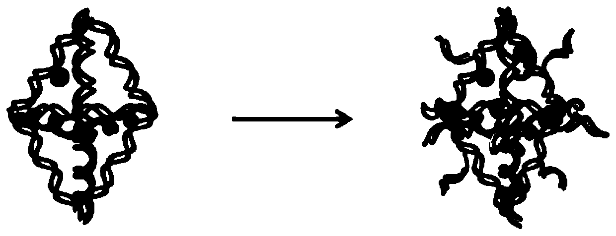
Nuclear magnetism-optical dual-mode imaging biconical DNA nanoprobe, and preparation method and application thereof
A dual-mode imaging and nanoprobe technology, applied in the field of nanomedicine, can solve the problems of increasing toxicity, difficulty in early diagnosis of tumors, and improving contrast agents.
- Summary
- Abstract
- Description
- Claims
- Application Information
AI Technical Summary
Problems solved by technology
Method used
Image


Examples
Embodiment 1
[0101] Example 1 Preparation of biconical DNA nanoprobes for NMR-optical dual-modal imaging
[0102] In this embodiment, the following method is used to prepare the NMR-optical dual-mode imaging biconical DNA nanoprobe, including the following steps:
[0103] 1. Preparation of DNA-DOTA(Gd) MRI contrast agent
[0104] The nuclear magnetic resonance imaging contrast agent is prepared by two-step organic chemical reactions. The specific steps are as follows: the first step, the 5' end amino modification, the 3' end Cy3 modified single-stranded DNA was dissolved in deionized water, and the concentration was adjusted to 200 μM; DOTA-NCS was dissolved in 80% DMSO, and the concentration was adjusted to 200 mM; Strand DNA and DOTA-NCS were mixed in 0.1M HEPES (pH=9, containing 300mM MgCl 2 ) buffer solution, add a magnet to stir, and react in a water bath at 37°C for 24h. This step reaction is the coupling reaction of amino group and isothiocyanate. In the second step, the obtaine...
Embodiment 2
[0126] Example 2 Preparation of biconical DNA nanoprobes for NMR-optical dual-modal imaging
[0127] The difference between this example and Example 1 is that the nuclear magnetic resonance imaging contrast agent selected in the process of synthesizing the nuclear magnetic-optical dual-modal imaging biconical DNA nanoprobe is DNA-DTPA (Gd), that is, DTPA and gadolinium are selected. Chelation. In addition, the selection of other raw materials, the preparation method and the reaction conditions are the same as those in Example 1, so as to prepare a biconical DNA nanoprobe for NMR-optical dual-mode imaging.
Embodiment 3
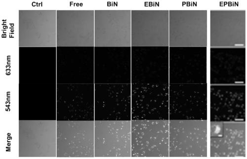
[0128] Example 3 Live Cell Uptake of Biconical DNA Nanoprobes by NMR-Optical Dual Mode Imaging
[0129] In this embodiment, the following methods are used to investigate the uptake of living cells by NMR-optical dual-modal imaging biconical DNA nanoprobes, as follows:
[0130] Due to the limitation of the excitation wavelength of the laser confocal scanning microscope (CLSM), the Dylight800 fluorescent molecule modified on the DNA probe was replaced by Cy5.5. MDA-MB-231 cells were cultured in a medium containing 10% fetal bovine serum and 1% double antibody (the double antibody was a mixture containing penicillin (10,000IU) and streptomycin (10,000μg / mL) at 100 times the working concentration) High-glucose DMEM complete medium, the culture environment is 37 ° C and 5% CO 2 . When the cells grow to a confluence of about 80%, the cells are digested and re-seeded in 35mm 2 In a confocal small dish, culture overnight for 12 hours. Replace the culture medium with DNA-DOTA (Gd),...
PUM
 Abstract
Description
Claims
Application Information
 - R&D
- Intellectual Property
- Life Sciences
- Materials
- Tech Scout
- Unparalleled Data Quality
- Higher Quality Content
- 60% Fewer Hallucinations
Browse by: Latest US Patents, China's latest patents, Technical Efficacy Thesaurus, Application Domain, Technology Topic, Popular Technical Reports.
© 2025 PatSnap. All rights reserved.Legal|Privacy policy|Modern Slavery Act Transparency Statement|Sitemap|About US| Contact US: help@patsnap.com