Ultra-low-field nuclear magnetic resonance imaging device applied to cerebral stroke diagnosis

An MRI, ultra-low field technology, applied in the direction of magnetic resonance measurement, diagnosis, application, etc., can solve the problems of cumbersome radiation protection equipment, limited application scenarios, and infrequent patients.

Inactive Publication Date: 2019-06-25
CHONGQING UNIV IND TECH RES INST
View PDF8 Cites 2 Cited by
  • Summary
  • Abstract
  • Description
  • Claims
  • Application Information

AI Technical Summary

Problems solved by technology

There is ionizing radiation in the CT imaging process, so patients cannot frequently undergo CT imaging. In addition, the CT imaging system has heavy radiation protection equipment. Although a vehicle-mounted CT has also been developed, the system is modified on the basis of a heavy truck. The cost is high and the application scenarios are limited; although there is no ionizing radiation in the imaging process of the traditional MRI system, due to its huge size, excessive weight, and complex superconducting maintenance technology, the imaging cost is high and the application scenarios are limited. Subject to greater restrictions

Method used


Image

  • Ultra-low-field nuclear magnetic resonance imaging device applied to cerebral stroke diagnosis
  • Ultra-low-field nuclear magnetic resonance imaging device applied to cerebral stroke diagnosis
  • Ultra-low-field nuclear magnetic resonance imaging device applied to cerebral stroke diagnosis

Examples


Embodiment 1

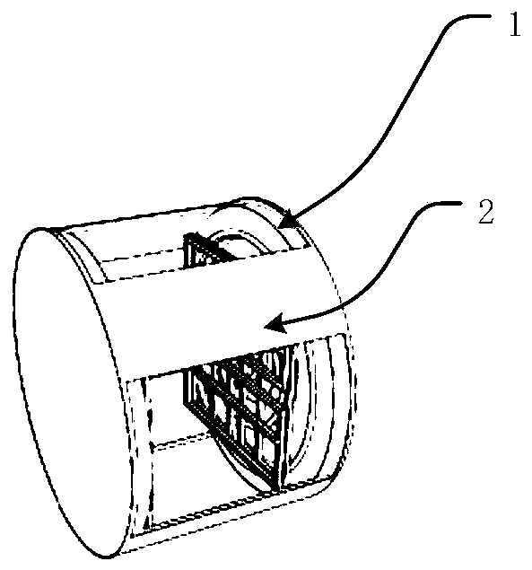
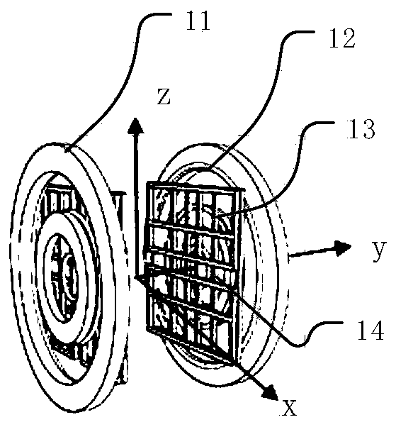
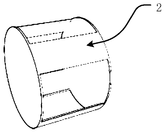
[0051] The present invention relates to an ultra-low-field nuclear magnetic resonance imaging device applied to the diagnosis of cerebral apoplexy. This embodiment relates to a disk-shaped ultra-low-field nuclear magnetic resonance imaging device, which includes a magnet system 1 and a supporting structure 2. The magnet system 1 for producing a spherical target area 3 at the geometric center of the magnet system 1, the support structure 2 is arranged outside the magnet system 1 for supporting the magnet system 1; the magnet system 1 comprises Main magnet 1 and gradient coils.

[0052] Optionally, the main magnet 1 includes two first magnets that are oppositely arranged, and a single first magnet is composed of several concentrically arranged circular coils; the gradient coils include a first gradient coil group 12 , the second gradient coil group 13 and the third gradient coil group; the first gradient coil group 12 includes two first gradient coils that are oppositely arrange...

Embodiment 2

[0058] This embodiment involves a saddle-shaped ultra-low-field nuclear magnetic resonance imaging device. The difference from Embodiment 1 is that the single first magnet is composed of several concentric hollow rectangular coils, and the single first magnet Both ends are bent towards another first magnet; both ends of a single first gradient coil are bent towards another first gradient coil; both ends of a single second gradient coil are bent towards another second gradient The direction of the coil is bent; both ends of a single third gradient coil are bent towards another third gradient coil.

[0059] In this embodiment, the main magnet 1 is made of copper wire, and its current direction is marked by the arrow in the figure. Only two pairs of coils in the first magnet are shown in the figure. According to the requirements of the main magnetic field strength and uniformity The number, size and relative position of the coils can be adjusted. The gradient magnetic field is ge...


PUM


Abstract

The present invention belongs to the field of magnetic resonance imaging devices, and relates to an ultra-low-field nuclear magnetic resonance imaging device applied to cerebral stroke diagnosis. Thedevice comprises a magnet system and a support structure, the magnet system is configured to generate a spherical target region, the target region is located at the geometric center of the magnet system, the support structure is arranged at the outer portion of the magnet system for supporting the magnet system; and the magnet system comprises a main magnet and a gradient coil. The ultra-low-fieldnuclear magnetic resonance imaging device can perform image monitoring for a brain for a long time, is convenient to move, light in weight, small in volume and compact in structure, can meet the clinic demands and can be applied to auxiliary clinical diagnosis.

Description

technical field [0001] The invention belongs to the field of nuclear magnetic resonance equipment, and relates to an ultra-low field nuclear magnetic resonance imaging device applied to the diagnosis of cerebral apoplexy. Background technique [0002] Stroke, also known as "stroke" or "cerebrovascular accident", is an acute cerebrovascular disease. Surveys show that stroke has become the number one cause of death in my country and the leading cause of disability among Chinese adults. Stroke has the characteristics of high morbidity, high mortality and high disability rate. After a stroke occurs, the treatment time has a great influence on the prognosis. If it is treated in time, the patient's prognosis will be good. If the treatment time is missed, the prognosis will be poor. The annual incidence of stroke in my country is increasing year by year, and it is estimated that the number of strokes will reach 3.7 million in 2020. At present, clinical diagnosis of early stroke d...

Claims


Application Information

Patent Timeline
IPC IPC(8): G01R33/38G01R33/385A61B5/055
Inventor 徐征沈晟王洪一贺玉成何为
Owner CHONGQING UNIV IND TECH RES INST
Why Eureka
Try Eureka