Potential-resolved electrochemiluminescence-based antigen detection method

A technology of antigen detection and electrochemistry, which is applied in the direction of chemiluminescence/bioluminescence, and analysis through chemical reaction of materials, can solve the problems of complex modulation, achieve high sensitivity, improve consumption, and fast analysis speed

Inactive Publication Date: 2019-03-15
ZHEJIANG UNIV
View PDF7 Cites 3 Cited by
  • Summary
  • Abstract
  • Description
  • Claims
  • Application Information

AI Technical Summary

Problems solved by technology

However, the modulation of the excitation process of electrochemiluminescence is more complicated, and the difficulty lies in the selection of the luminescent body and the control of the excitation potential.

Method used


Image

  • Potential-resolved electrochemiluminescence-based antigen detection method
  • Potential-resolved electrochemiluminescence-based antigen detection method
  • Potential-resolved electrochemiluminescence-based antigen detection method

Examples


Embodiment 1

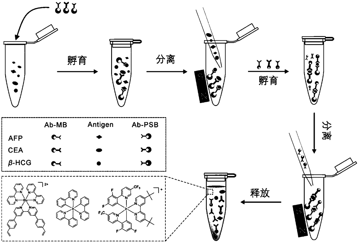
[0068] Such as image 3 As shown, alpha-fetoprotein (AFP), carcinoembryonic antigen (CEA) and human chorionic gonadotropin (β-HCG) were detected simultaneously.

[0069] The simulated sample for simultaneous detection of multiple indicators is the PBS solution containing CEA, AFP and β-HCG. According to the clinical detection values ​​of tumor markers, the final concentrations of CEA, AFP and β-HCG in the detection system were adjusted to 5 ng / mL, 25 ng / mL and 5 mIU / mL, respectively. The process of simultaneous detection of multiple tumor markers mainly includes steps such as incubation, magnetic separation, washing, and detection. The specific operation is as follows:

[0070] (1) Add 100 μL of magnetic bead solutions labeled with three kinds of capture antibodies (the concentration of magnetic beads is 1 mg / mL) into 300 μL of PBS mock sample, and incubate for 30 min in a constant temperature shaker at 37°C. After incubation, place the centrifuge tube on the magnetic separ...

Embodiment 2

[0077] Add any pair of detection antibody-labeled polymer microspheres and capture antibody-labeled magnetic beads into the simulated sample to investigate the selective detection performance of a single indicator in multi-index samples.

[0078] Taking the selective detection of AFP as an example, take 100μL containing 1mg / mL Ab (AFP) -MB PBS buffer solution and 200 μL 0.5% BSA / PBS solution were added to 300 μL of the above sample, and incubated at 37° C. for 30 min. Separate / wash, repeat 3 times; then add 300 μL 0.5% BSA / PBS solution, 25 μL 1mg / mL Ab (AFP) -Ru@PSB and 50 μL of 0.5% BSA / PBS solution, mix well, and incubate at 37° C. for 30 minutes. Separation / washing was repeated 5 times. After vacuum drying, 100 μL of acetonitrile solution was added and ultrasonically swelled. When detecting CEA and β-HCG, the concentration of probe molecules added was 10 mg / mL, and other operation steps were consistent with the above-mentioned procedures.

[0079] Figure 6 Shown are th...


PUM

PropertyMeasurementUnit
emission peakaaaaaaaaaa

Abstract

The invention discloses a potential-resolved electrochemiluminescence-based antigen detection method. The method comprises the steps of: preparing an electrochemiluminescent probe; doping the electrochemiluminescent probe into a polymer microsphere; marking a detection antibody onto the surface of the polymer microsphere so as to obtain a double-encoded polymer microsphere double of the detectionantibody and the electrochemiluminescent probe; covalently marking a capture antibody to the surface of a magnetic microsphere so as to obtain an immunomagnetic microsphere marked by the capture antibody; placing the double-encoded polymer microsphere and the immunomagnetic microsphere into to-be-detected solution, carrying out magnetic separation, and releasing an electrochemical probe by swelling the polymer microsphere; acquiring electrochemical luminescence signals of the electrochemical probe at different potentials to identify the detected antigen. The antigen detection method provided by the invention is capable of improving the defect that the single-component electrochemiluminescence immunoassay is long in time and low in flux, and realizing rapid, high-throughput and high-sensitivity joint detection of various tumor markers, thereby being suitable for developing kits for target tumors for large-scale early screening of tumors.

Description

technical field [0001] The invention belongs to the field of simultaneous identification and detection of tumor markers, in particular to an antigen detection method based on potential-resolving electrochemiluminescence. Background technique [0002] Routine imaging examinations are insufficient in the early diagnosis of tumors, and tissue biopsy techniques are more invasive to patients and are not suitable for prognosis judgment and efficacy monitoring. Tumor markers are a class of substances synthesized and secreted by tumor cells through gene expression, or produced by the body in response to tumors, and play an important role in early tumor screening, prognosis judgment, and efficacy monitoring. Therefore, it is necessary to develop highly sensitive immunosensors for the detection of tumor markers. Most of the reported immunosensors are used for single tumor marker detection. However, tumor markers are not organ-specific, and the sensitivity or specificity of a single ...

Claims


Application Information

Patent Timeline
Patent Type & Authority Applications(China)
IPC IPC(8): G01N21/76
CPCG01N21/76
Inventor 苏彬郭维亮丁昊刘燕欢
Owner ZHEJIANG UNIV
Why Eureka
Try Eureka