
Auxiliary device for vitreous cavity injection

An auxiliary device and vitreous cavity technology, applied in the field of medical devices, can solve the problems of poor patient compliance, time-consuming, and no uniform standard for the location and depth of injections

Active Publication Date: 2019-01-15
LONGHUA HOSPITAL SHANGHAI UNIV OF TRADITIONAL CHINESE MEDICINE
View PDF8 Cites 3 Cited by
  • Summary
  • Abstract
  • Description
  • Claims
  • Application Information

AI Technical Summary

Problems solved by technology

[0003] 1. Systemic administration: due to the effect of the inner and outer blood-retinal barriers, the amount of drug absorbed into the intraocular tissues such as vitreous body and retina is very small (the drug concentration in the vitreous body is often less than 10% of the drug concentration in plasma), up to If the drug concentration is less than the effective drug concentration, increasing the dosage can partially increase the drug concentration in the retinal tissue of the eye, but it may also cause serious systemic side effects
[0004] 2. Ocular surface administration (mainly eye drops): affected by the rapid loss of drugs on the ocular surface and the aqueous humor barrier, it is difficult for the drug to reach an effective drug concentration in the retinal tissue after instillation of conventional eye drops
[0005] 3. Peribulbar injection: Compared with systemic administration and ocular surface administration, this route of administration is easier to reach the intraocular tissue, and it is less traumatic than vitreous injection, but its curative effect is largely affected by how it is delivered to the retina, The limitation of the difficult problem of transporting effective doses of drugs in the posterior segment of the eye such as the choroid, in order to maintain the effective drug concentration, repeated injections must be performed, and the patient's compliance is poor
[0009] However, during intravitreal injection, the drug needs to be delivered to the vitreous by injecting the needle at 3.5 or 4 mm behind the limbus (here is the pars plana of the ciliary body without retinal tissue and depending on the state of the lens of the eye to be injected). To avoid complications such as retinal breaks and detachment, each operation needs to be accurately measured with a ruler. For inexperienced doctors, it is time-consuming and inefficient
Secondly, the injection needle needs to be inserted into the center of the eyeball. Improper operation can damage the lens, retina and other adjacent tissues. For inexperienced doctors, it may cause serious complications
In addition, there is no uniform standard for the position and depth of intravitreal injection, and the operation is basically done by referring to the instructions of each product.

Method used


Image

  • Auxiliary device for vitreous cavity injection
  • Auxiliary device for vitreous cavity injection
  • Auxiliary device for vitreous cavity injection

Examples


Embodiment Construction

[0022] The present invention will be further elaborated below in conjunction with the accompanying drawings and specific embodiments. These examples should be understood as only for illustrating the present invention but not for limiting the protection scope of the present invention. After reading the contents of the present invention, those skilled in the art can make various changes or modifications to the present invention, and these equivalent changes and modifications also fall within the scope defined by the claims of the present invention.

[0023] The parameters of the auxiliary device for intravitreal injection in the following examples of the present invention are all based on an eyeball diameter of 24mm.

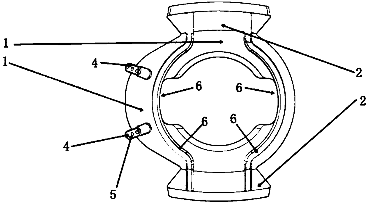
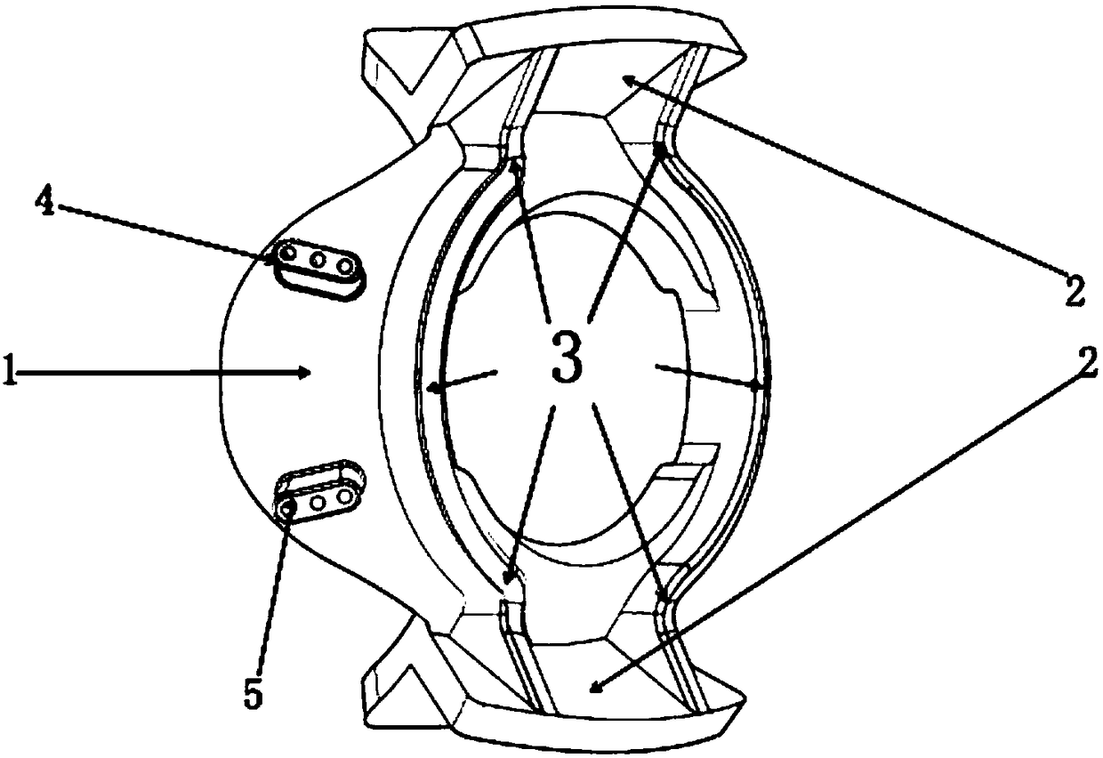
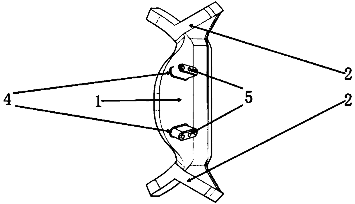
[0024] Such as Figure 1-Figure 5 As shown, the newly designed auxiliary device for intravitreal injection provided by the preferred embodiment of the present invention consists of a fixed ring 1, an eyelid support 2 and a support beam 3 connecting the two, where...


PUM

PropertyMeasurementUnit
Thicknessaaaaaaaaaa
Outer diameteraaaaaaaaaa
The inside diameter ofaaaaaaaaaa

Abstract

The invention discloses an auxiliary device for vitreous cavity injection, which is composed of a fixing ring, an eyelid support and a support beam connecting the eyelid and the eyelid. The drug can be injected into the vitreous cavity safely and effectively according to the size of the eyeball, the position and depth of the drug administration are relatively fixed, the use is convenient, the operation is simple, the operation risk is reduced, the operation efficiency is improved, and the requirement of ergonomics is met.

Description

technical field [0001] The invention belongs to the technical field of medical devices, and in particular relates to an auxiliary device for vitreous cavity injection (or vitreous cavity puncture and liquid extraction). Background technique [0002] At present, with the arrival of aging, the incidence of diseases in the posterior segment of the eye is increasing day by day, which is also one of the main causes of vision loss. For example: epidemiological survey results show that age-related macular degeneration (age-related macular degeneration, AMD) is the leading cause of blindness in Western countries over the age of 50, and the incidence rate in Asia is also increasing year by year; diabetic retinopathy ( Diabetic retinopathy, DR) has always been the most important blinding eye disease among working-age people in developed countries. Two population-based epidemiological studies organized by Beijing Tongren Eye Center in recent years found that the prevalence of DR among ...

Claims


Application Information

Patent Timeline
IPC IPC(8): A61F9/007A61F9/00
CPCA61F9/0026A61F9/00736
Inventor 王大虎刘新泉单亚洲
Owner LONGHUA HOSPITAL SHANGHAI UNIV OF TRADITIONAL CHINESE MEDICINE
Learn More