
Patsnap Eureka
For R&D, Patsnap Eureka makes reading and utilizing patents & technical documents easy.
Patsnap Eureka AIR
Designed for self-driven R&D workflows. Generate viable solutions, solve complex R&D challenges, empower your innovation with AI.

Patsnap Eureka Materials
Designed for material experts only. Revolutionize your material R&D, from search, analyze, to developing new materials.

TechResearch
Generate reliable direction feasibility study reports for your R&D in just a few steps.

TechSeek
Discover and master advanced knowledge NOW. Basics, ideas, possibilities, all at once.

TechMind
As an expert in R&D Theories, TechMind can generates customized viable solutions instantly.

TechRisk
Analyze your overall solution with one click, know your potential R&D risks in advance.

TechMonitor
Get weekly tech updates, stay abreast of the latest tech innovations and key insights.
Kit and method for detecting EBV infection in trace biological sample of eye
A biological sample and ocular technology, applied in biochemical equipment and methods, microbiological measurement/inspection, DNA/RNA fragments, etc., can solve the problem of lack of high-sensitivity detection methods for virus infection in eye tissue, easy misdiagnosis and inaccurate diagnosis Reflect the real cause of the eye and other problems
- Summary
- Abstract
- Description
- Claims
- Application Information
AI Technical Summary
Problems solved by technology
Method used
Image

Examples
Embodiment 1
[0121] Example 1. Method for extracting DNA from test specimens of eye microfluidics
[0122] Material:
[0123] Specimens to be tested: collected from admitted patients, tear fluid, aqueous humor 2, and vitreous.
[0124] (1) Put the collected sample to be tested into a container, add 10-30 μl proteinase K, 100-300 μl lysis buffer AL, and treat at 56°C for more than 10 minutes;
[0125] (2) Add the same volume of absolute ethanol as the lysis buffer, shake and mix for 10-20s, and centrifuge briefly;
[0126] (3) Put the liquid obtained in step (2) into a spin column, put it into a 2ml collection tube, and centrifuge at 6000-9000rpm for 0.5-2min;
[0127] If the sample volume > 140 μl, repeat step (3);
[0128] (4) Add 300-600μl elution buffer 1, 6000-9000rpm, 0.5-2min, discard the filtrate and collection tube, and replace with a new collection tube;
[0129] (5) Add 300-600μl Elution Buffer 2, centrifuge at 10000-15000rpm for 1-5min, discard the filtrate and collection tu...
Embodiment 2
[0137] Example 2. Method for extracting DNA from eye trace solid specimens to be tested
[0138] Material:
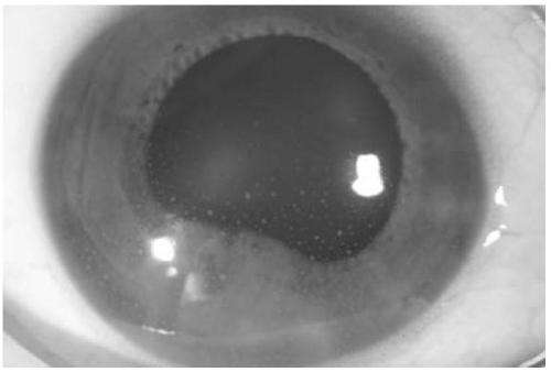
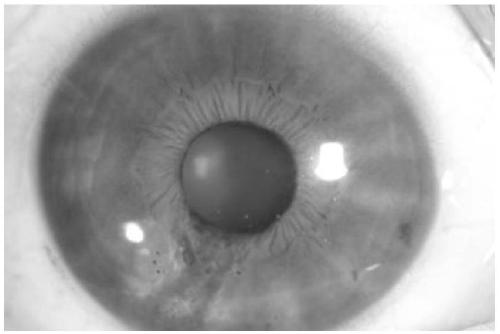
[0139] Specimens to be tested: collected from admitted patients, including retina, corneal endothelium, pterygium, conjunctiva, iris, and eye tumors, with a collection volume of 1×1mm;
[0140] step:
[0141] (1) Put the collected sample to be tested into a container, add 10-30 μl proteinase K, 100-300 μl lysis buffer AL, and treat at 56°C for 6-12 hours;
[0142] (2) Add the same volume of absolute ethanol as the lysis buffer, shake and mix for 10-20s, and centrifuge briefly;
[0143] (3) Put the liquid obtained in step (2) into a spin column, put it into a 2ml collection tube, and centrifuge at 6000-9000rpm for 0.5-2min;
[0144] (4) Add 300-600μl elution buffer 1, 6000-9000rpm, 0.5-2min, discard the filtrate and collection tube, and replace with a new collection tube;
[0145] (5) Add 300-600μl Elution Buffer 2, centrifuge at 10000-15000rpm for 1-5min, discard th...
Embodiment 3
[0153] Example 3. Kit for detection of ocular EBV infection by ocular microsamples
[0154] DNA extraction reagent set:
[0155] Lysis buffer: Tris-saturated phenol with 10% SDS,
[0156] Elution buffer 1: a mixture of saturated phenol: chloroform: isoamyl alcohol with a volume ratio of 25:24:1;
[0157] Elution buffer 2: absolute ethanol,
[0158] Elution buffer 3: pH 8.0, 10 mmol / L Tris-HCl solution containing 1 mmol / LEDTA.
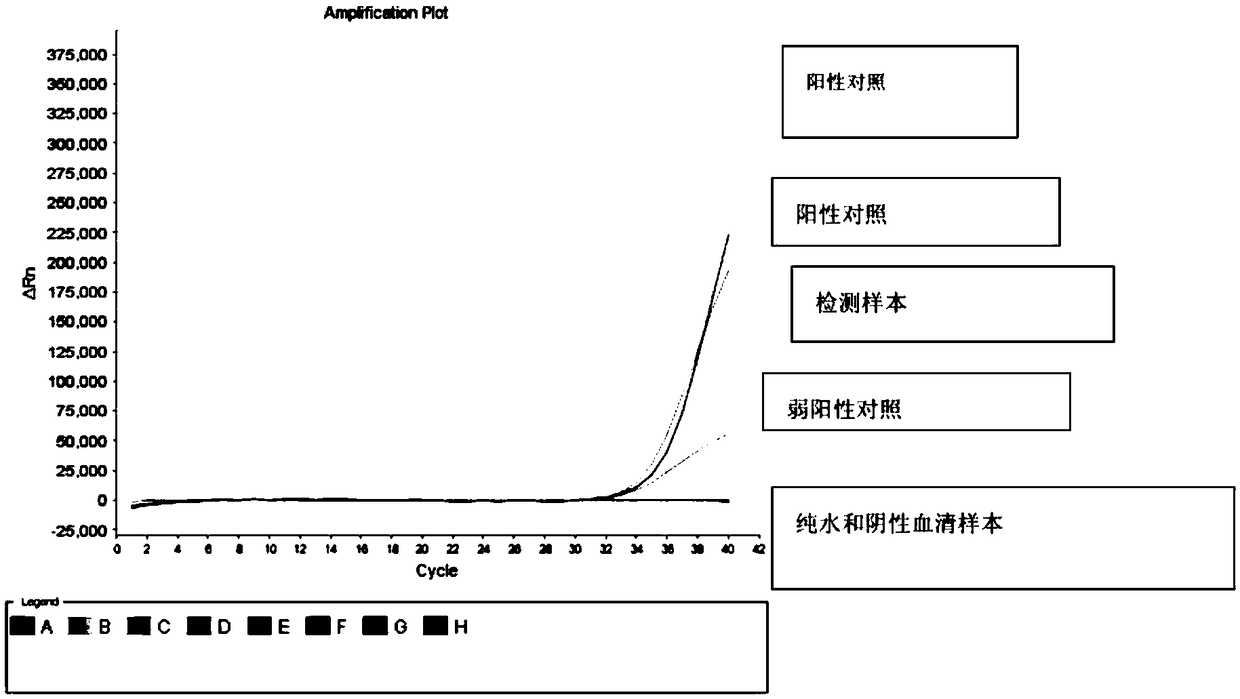
[0159] Specific primers and probes for PCR amplification
[0160] Primer-F: CAACGTGTGCCTCTTTCTTCAT
[0161] Primer-R: ACCACCAACGGGACTGTCATG
[0162] Probe: CAACGGGACTGTCATGGAAATT
PUM
| Property | Measurement | Unit |
|---|---|---|
| diameter | aaaaa | aaaaa |
Abstract
Description
Claims
Application Information
 - R&D Engineer
- R&D Manager
- IP Professional
- Industry Leading Data Capabilities
- Powerful AI technology
- Patent DNA Extraction
Browse by: Latest US Patents, China's latest patents, Technical Efficacy Thesaurus, Application Domain, Technology Topic, Popular Technical Reports.
© 2024 PatSnap. All rights reserved.Legal|Privacy policy|Modern Slavery Act Transparency Statement|Sitemap|About US| Contact US: help@patsnap.com