Influenza luciferase reporter virus-based animal model building method and application
An animal model, luciferase technology, applied in the biological field, achieves the effects of saving feeding time, saving the number of experimental animals, and reducing costs
- Summary
- Abstract
- Description
- Claims
- Application Information
AI Technical Summary
Problems solved by technology
Method used
Image
Examples
Embodiment 1
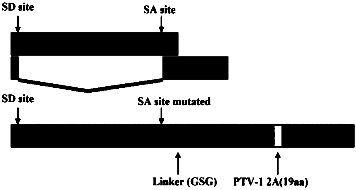
[0045] A reverse genetics system was used to rescue and amplify a recombinant influenza virus PR8-NS1-Gluc carrying the Gaussia luciferase gene with a replication ability similar to that of the wild type. The steps are as follows:
[0046] Eight plasmids (0.5 μg / plasmid) required by the influenza reverse genetics system (provided by Dr. Balaji Manicassamy, University of Chicago) carrying the Gaussia luciferase gene were co-transfected into co-cultured 293T / MDCK cells, and after 12 hours of transfection, The culture medium was discarded and replaced with Opti-MEM medium containing 2 μg / ml TPCK-Trypsin (purchased from Sigma, USA). After culturing for 72 hours, the supernatant was collected and inoculated into eight-day-old chicken embryos. After 48 hours, the allantoic fluid of chicken embryos was collected, which was the required recombinant influenza virus.
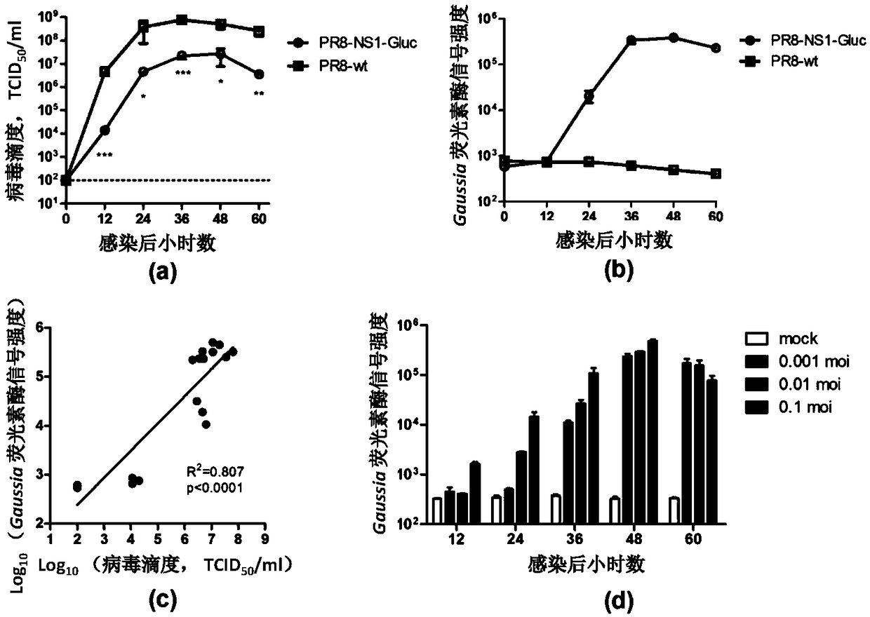
[0047] The amplified virus and the wild-type virus were infected with MDCK cells at 0.01 MOI respectively, and samples...
Embodiment 2
[0049] Determination of virulence of influenza luciferase reporter virus PR8-NS1-Gluc in mice. The steps are:
[0050] (1) Dilute the recombinant virus PR8-NS1-Gluc virus liquid with PBS to contain 10 2 -10 6 TCID 50 virus.
[0051] (2) 4-6 weeks old female BALB / c mice were divided into random groups, 10 in each group.
[0052] (3) Mice were inhaled and anesthetized with isoflurane, and 30 μl of serially diluted virus solution was taken to instill nasally to infect the mice.
[0053] (4) The body weight change and survival of the mice were monitored daily for 14 days.
[0054] as attached image 3 , using the recombinant virus to infect 4-6 week-old female BALB / c mice, the mice showed obvious weight loss and infection dose-dependent death. This result shows that the obtained recombinant reporter virus can effectively infect mice and has high virulence, and can be used to establish mouse models of influenza sublethal infection and lethal dose infection.
Embodiment 3
[0056] Detection of influenza luciferase reporter virus PR8-NS1-Gluc specific for infected tissues in mice. The specific steps are:
[0057] (1) Dilute the virus liquid with PBS to contain 1000TCID per 30μl PBS 50 virus.
[0058] (2) Five 4-6 week old female BALB / c mice were anesthetized by breathing with isoflurane, and 30 μl of diluted virus liquid was taken to infect the mice nasally.
[0059] (3) Six days after infection, the mice were sacrificed and dissected. For 3 of them, lung tissue, liver tissue, spleen, etc. were collected, respectively, and 500 μl of PBS were added for homogenization treatment. For the other two, lung tissues were taken and fixed in formalin. In each experiment, tissues from healthy mice were used for the same treatment as controls.
[0060] (5) Take 50 μl of lung tissue homogenate and dilute to 1 ml with PBS. Take 20 μl and use BioLux Gaussia Luciferase Assay Kit (NEB) to measure luciferase activity according to the instructions.
[0061] (...
PUM
 Abstract
Description
Claims
Application Information
 - R&D Engineer
- R&D Manager
- IP Professional
- Industry Leading Data Capabilities
- Powerful AI technology
- Patent DNA Extraction
Browse by: Latest US Patents, China's latest patents, Technical Efficacy Thesaurus, Application Domain, Technology Topic, Popular Technical Reports.
© 2024 PatSnap. All rights reserved.Legal|Privacy policy|Modern Slavery Act Transparency Statement|Sitemap|About US| Contact US: help@patsnap.com


