Pepsinogen I (PGI) detection kit and detection method thereof
A pepsinogen and detection kit technology, applied in the field of biomedicine, can solve the problems of expensive equipment, poor stability, etc., and achieve the effects of reducing the amount of detection samples, increasing stability, and improving work efficiency
- Summary
- Abstract
- Description
- Claims
- Application Information
AI Technical Summary
Problems solved by technology
Method used
Image
Examples
Embodiment 1
[0033] Pepsinogen I (PGI) detection kit and detection method thereof, adding glycerol stabilizer:
[0034] R 1 : Phosphate buffer 100mmoL / L, Proclinc 3000.5g / L, polyethylene glycol 600010g / L, Triton 1001g / L, TRIS 10g / L;
[0035] R 2 : Phosphate buffer 100mmoL / L, latex particle anti-human PGI antibody-conjugated emulsion (anti-human pepsinogen I (mouse) monoclonal antibody sensitive emulsion solution 1.3mg / mL), Proclinc 3000.5g / L, calf serum Albumin (BSA) 10g / L, glycerol 90g / L;
[0036] Wherein, the preparation method of latex particle anti-human PGI antibody binding emulsion (anti-human pepsinogen I (mouse) monoclonal antibody sensitive emulsion solution) is:
[0037] Step 1: Take 2mL of carboxylated latex microspheres (150nm, 5% w / v) and add phosphate buffer to 4ml;
[0038]Step 2: Add 50ml 0.2g / L of N-hydroxysuccinimide (NHS) and 50ml 0.2g / L of 1-(3-dimethylaminopropyl)-3-ethylcarbodiimide hydrochloride Salt (EDC) for activation, stir well and incubate at 37°C for 20min...
Embodiment 2
[0043] Pepsinogen I (PGI) detection kit and its detection method, without glycerol:
[0044] R 1 : Phosphate buffer 100mmoL / L, Proclinc 3000.5g / L, polyethylene glycol 600010g / L, Triton 1001g / L, TRIS 10g / L;
[0045] R 2 : Phosphate buffer 100mmoL / L, latex particle anti-human PGI antibody conjugated emulsion (anti-human pepsinogen I (mouse) monoclonal antibody sensitive emulsion solution: 1.3mg / mL), Proclinc 3000.5g / L, calf Serum albumin (BSA) 10g / L;
[0046] Wherein, the preparation method of latex particle anti-human PGI antibody binding emulsion (anti-human pepsinogen I (mouse) monoclonal antibody sensitive emulsion solution) is:
[0047] Step 1: Take 2mL of carboxylated latex microspheres (150nm, 5% w / v) and add phosphate buffer to 4ml;
[0048] Step 2: Add 50ml 0.2g / L of N-hydroxysuccinimide (NHS) and 50ml 0.2g / L of 1-(3-dimethylaminopropyl)-3-ethylcarbodiimide hydrochloride Salt (EDC) for activation, stir well and incubate at 37°C for 20min;
[0049] Step 3: Add 3 mL...
Embodiment 3
[0053] Embodiment three: the accuracy analysis of reagent of the present invention and method:
[0054] Test instrument: Hitachi 7170 automatic biochemical analyzer;
[0055] Test samples: 40 random serum samples and one PGI serum sample (target value 59.75ng / mL)
[0056] Control kit: Protease I (PGI) detection kit (latex immunoturbidimetric method) from a manufacturer approved by the State Food and Drug Administration (including reagents R1 and R2, but the composition is different from the present invention, hereinafter referred to as the control Reagent);
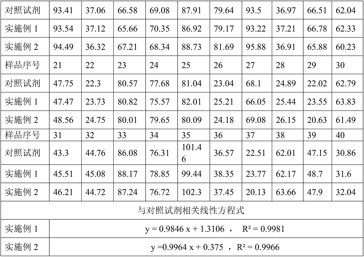
[0057] Using the reagents of Examples 1 and 2 and the comparative reagents, respectively, by their respective detection methods, 40 serum samples and a PGI serum sample (target value of 59.75 ng / mL) were simultaneously measured, and the results are shown in Table 1 and Table 2 Shown:
[0058] Table 1 The results of different detection reagents measuring 40 cases of random serum samples (unit: ng / mL)
[0059]
[006...
PUM
 Abstract
Description
Claims
Application Information
 - R&D
- Intellectual Property
- Life Sciences
- Materials
- Tech Scout
- Unparalleled Data Quality
- Higher Quality Content
- 60% Fewer Hallucinations
Browse by: Latest US Patents, China's latest patents, Technical Efficacy Thesaurus, Application Domain, Technology Topic, Popular Technical Reports.
© 2025 PatSnap. All rights reserved.Legal|Privacy policy|Modern Slavery Act Transparency Statement|Sitemap|About US| Contact US: help@patsnap.com


