Method for observing dynamic distribution of tumor cell exosome and exosome internal miRNA in recipient cells

A tumor cell and receptor cell technology, applied in the field of biophotonics, can solve the problem of inability to observe exosomes in detail, and achieve the effects of many switching times, small bright state duty cycle, and good photostability

Inactive Publication Date: 2018-06-22
SOUTHEAST UNIV
View PDF2 Cites 7 Cited by
  • Summary
  • Abstract
  • Description
  • Claims
  • Application Information

AI Technical Summary

Problems solved by technology

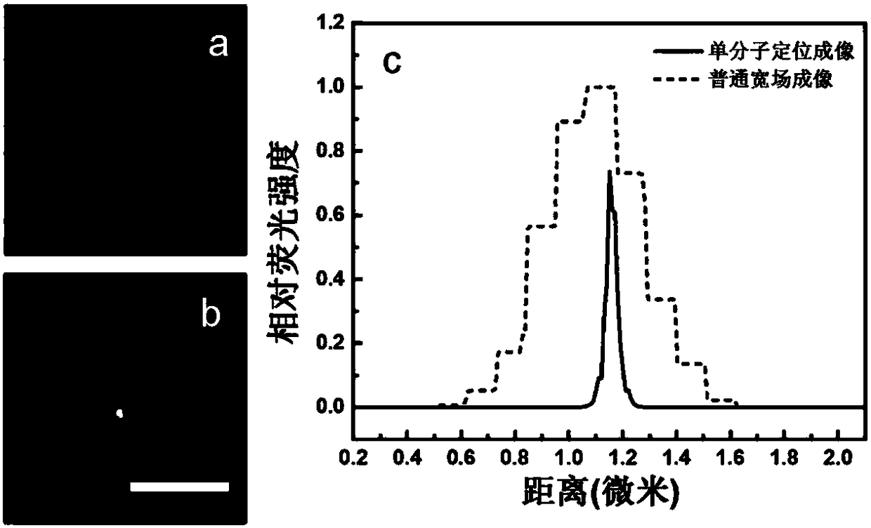
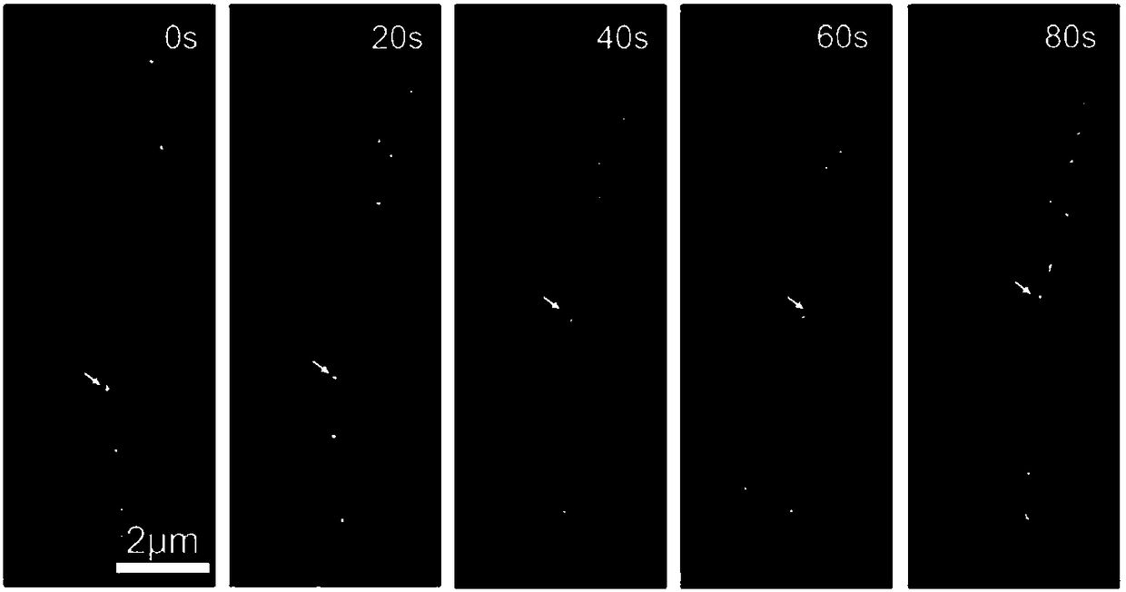
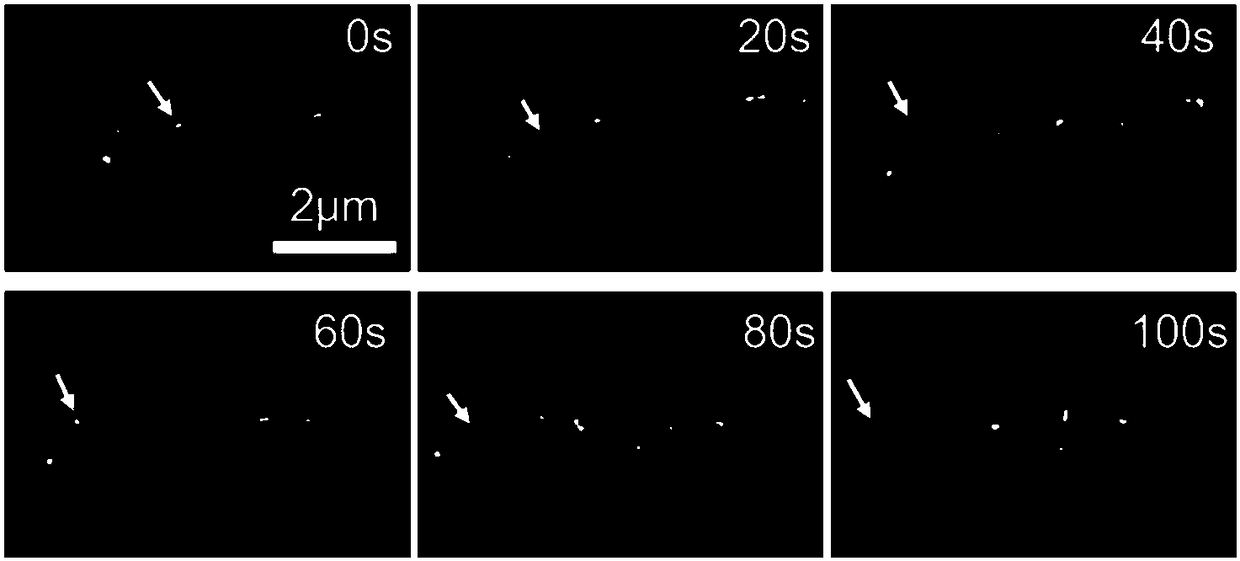
[0004] In the above background, the present invention proposes to use super-resolution optical imaging technology based on single-molecule localization to observe the dynamic distribution of tumor cell exosomes and miRNAs inside exosomes in recipient cells, which solves the problem of diffraction limit in the prior art. Limiting the problem of not being able to observe exosomes in detail, it provides a new technical means for studying the mechanism of exosome-mediated cancer metastasis and the treatment of cancer metastasis

Method used


Image

  • Method for observing dynamic distribution of tumor cell exosome and exosome internal miRNA in recipient cells
  • Method for observing dynamic distribution of tumor cell exosome and exosome internal miRNA in recipient cells
  • Method for observing dynamic distribution of tumor cell exosome and exosome internal miRNA in recipient cells

Examples


Embodiment 1

[0029] The PBS buffer involved in this example is pH=7.4, the concentration is 10mM PBS buffer; the receptor cell membrane dye involved is PKH67, and the reaction solution is Diluent C solution provided by sigma; the involved single molecule localization super-resolution The optical imaging technology is PALM technology, and the low-toxic imaging buffer involved is DMEM medium, which is equipped with 27mM ß-mercaptoethanol, 1wt% glucose, 0.1mg / mL glucose oxidase and 8μg / mL catalase The tumor cell exosomes involved are HeLa cell exosomes, and the recipient cells are PC12 cells; the internal miRNA involved tumor cell exosomes is mir-21, and its sequence is 3'AGUUGUAGUCAGACUAUUCGAU 5', the molecular beacon used The MB21 sequence is 5'Cy5-CTCTTTCAACATCAGTCTGATAAGCTAAAGAG-BHQ3 3', where Cy5 is a fluorescent molecule and BHQ3 is a quenching group. The exosomal membrane dye involved is DiI; the involved exosomal extract is the exosome extract included in the ExoQuick-TC Exosomes Preci...

Embodiment 2

[0039] The PBS buffer involved in this embodiment is pH=7.4, and the concentration is 10mM PBS buffer; the receptor cell membrane dye involved is DiO; the single molecule localization super-resolution optical imaging technology involved is PALM technology, which involves low-toxicity imaging The buffer is DMEM medium with 27mM ß-mercaptoethanol, 1wt% glucose, 0.1mg / mL glucose oxidase and 8μg / mL catalase; the tumor cell exosomes involved are HeLa extracellular Exosomes, the recipient cell is PC12 cells; the internal miRNA of tumor cell exosomes involved is mir-21, its sequence is 3'AGUUGUAGUCAGACUAUUCGAU 5', and the molecular beacon MB21 sequence used is 5'Alexa Fluor 647-CTCTTTCAACATCAGTCTGATAAGCTAAAGAG-BHQ3 3', where Alexa Fluor 647 is a fluorescent molecule and BHQ3 is a quenching group. The involved exosomal membrane dye is PKH26; the involved exosome extract is the exosome extract included in the ExoQuick-TC Exosomes Preciptation Solution exosome kit of SBI.

[0040] Step 1:...


PUM

PropertyMeasurementUnit
sizeaaaaaaaaaa

Abstract

The invention discloses a method for observing dynamic distribution of tumor cell exosome and exosome internal miRNA in recipient cells. The method comprises the following steps: after extracting thetumor cell exosome, conducting fluorescence labeling on a tumor cell exosome membrane and the exosome internal miRNA through an exosome membrane stain and a molecular beacon which is complementary with the exosome internal miRNA, and meanwhile, staining the recipient cells via a cell membrane stain; and implementing co-culture on the tumor cell exosome obtained from the fluorescence labeling and the stained recipient cells, adding an imaging buffer solution after an interaction of the tumor cell exosome and the recipient cells, and implementing dynamic tracing of exosome and miRNA in living cells through a super-resolution optical microscope. According to the method provided by the invention, the problem in the prior art that exosome cannot be carefully observed due to diffraction limit can be solved; and a novel technological means can be provided for researches on an exosome-mediated cancer metastasis mechanism and cancer metastasis and spread treatment.

Description

Technical field [0001] The present invention relates to the field of biophotonics, in particular to a method for observing the dynamic distribution of miRNA in tumor cell exocrine and exosomes in recipient cells. Background technique [0002] Exosomes are a type of vesicles secreted by cells, with a size of about 30-100 nm. The outer layer of this interesting nano-scale vesicle is a lipid membrane, on which specific signal molecules are expressed, and the inside contains biological molecules such as protein and RNA. Exosomes play a very important role in cell-cell communication and material exchange. Existing studies have shown that the interactions between exosomes and recipient cells are mainly divided into three types: 1) Exosomes bind to the receptor cell surface through exosomal adhesion molecules on the receptor cell surface; 2) Exosomes directly fuse with recipient cells; 3) Exosomes enter the recipient cells through endocytosis received by the receptor. Tumor cell exos...

Claims


Application Information

Patent Timeline
Patent Type & Authority Applications(China)
IPC IPC(8): C12Q1/6816G01N21/64
CPCC12Q1/6816G01N21/6486C12Q2563/107C12Q2525/207
Inventor 宗慎飞陈晨王著元崔一平
Owner SOUTHEAST UNIV
Why Eureka
Try Eureka