Method for inducing directional myocardial differentiation of iPSCs by adopting HCM myocardial cell culture fluid
A technique for culturing cardiomyocytes and cells, which is applied in the field of medicine and can solve problems such as different induction effects
- Summary
- Abstract
- Description
- Claims
- Application Information
AI Technical Summary
Problems solved by technology
Method used
Image

Examples
Embodiment 1
[0017] Example 1 Preparation of iPSCs
[0018] 1) Preparation of trophoblast layer (MEF): Add 0.1% (volume fraction) gelatin into a T25 culture flask, put it in a cell culture incubator at 37°C for 20 minutes, then suck it off, add 5-6 mL of MEF culture medium preheated at 37°C At the same time, the mouse embryonic fibroblasts (MEF) (purchased from the cell bank of the Chinese Academy of Sciences in Shanghai) were quickly taken out from the liquid nitrogen, placed in a 37°C water bath to melt quickly, and immediately wiped and frozen with 75% alcohol by volume fraction Transfer the cell suspension in the cryopreservation tube to a 15mL centrifuge tube containing MEF culture medium, centrifuge at 1000rpm for 5min, discard the supernatant, resuspend it, add it to a T25 culture bottle, and place it in a CO2 In a constant temperature incubator, iPSCs can be added to the feeder layer after 24 hours of culture;
[0019] 2) Cultivation and passage of iPSCs: The trophoblast obtained ...
Embodiment 2
[0020] Example 2 HCM cardiomyocyte culture medium induces iPSCs differentiation
[0021] HCM cardiomyocytes were resuspended in DMEM medium containing 15% FBS, 50 U / mL penicillin, and 10 μg / mL streptomycin, and inoculated at an appropriate cell density in T25 culture flasks for culture, and passaged for 1 to 2 days. Aspirate the culture medium before subculture for 3 days, and filter it through a 0.22 μm filter membrane to obtain the induction culture medium, that is, the HCM cardiomyocyte culture medium, and set it aside.
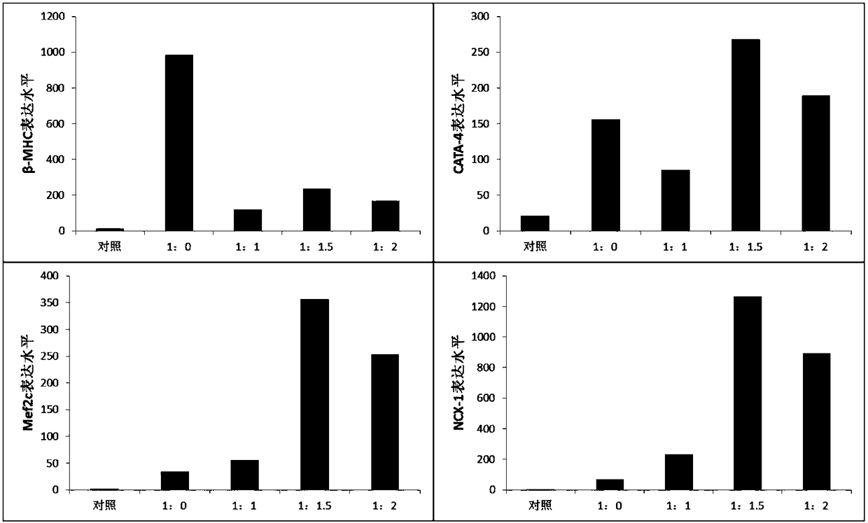
[0022] The iPSCs in the logarithmic phase of growth were taken, cultured with 100×106 cells / mL iPSCs hanging drop for 48 hours, and seeded in a six-well plate pretreated with 0.1% gelatin for 1 hour. The cell culture medium was mixed at a ratio of 1:0, 1:1, 1:1.5, and 1:2 to make a differentiation medium with 15% fetal bovine serum to cultivate iPSCs. Conventional untreated iPSCs served as the control group.
[0023] Conventional iPSCs cell culture mediu...
Embodiment 3
[0024] Example 3 qPCR detection of myocardial specific protein expression after iPSCs were induced and pretreated
[0025] The total RNA of the sample cells was extracted according to the TRIZOL operating instructions. The total RNA was reverse-transcribed with reverse transcriptase to obtain cDNA; it was amplified on a PCR machine, and the Ct values of the target gene and the internal reference gene were obtained according to the real-time quantitative amplification curve, and the relative quantitative value of the target gene expression was used for statistical analysis. The experiment was repeated three times.
[0026] RT-PCR detection of myocardial specific marker mRNA expression in induced pluripotent stem cell embryoid bodies, the results are as follows figure 1 As shown, the results showed that compared with the control group, among the four myocardial-specific markers of GATA-4, Mef2c, β-MHC and NCX-1, the pre-prepared conventional iPSCs cell culture medium with LIF...
PUM
 Abstract
Description
Claims
Application Information
 - R&D
- Intellectual Property
- Life Sciences
- Materials
- Tech Scout
- Unparalleled Data Quality
- Higher Quality Content
- 60% Fewer Hallucinations
Browse by: Latest US Patents, China's latest patents, Technical Efficacy Thesaurus, Application Domain, Technology Topic, Popular Technical Reports.
© 2025 PatSnap. All rights reserved.Legal|Privacy policy|Modern Slavery Act Transparency Statement|Sitemap|About US| Contact US: help@patsnap.com