
Visual three-dimensional imaging method for fetal body surface structure based on magnetic resonance scanning

A magnetic resonance scanning and three-dimensional imaging technology, which is applied in the field of medical imaging, can solve the problems of not being able to obtain a satisfactory three-dimensional image of the fetal body surface, lack of confirmation and support from other imaging examinations, and obtaining the structure of the fetal body surface, so as to achieve excellent image quality, The effect of intuitive image and broad application prospects

Inactive Publication Date: 2018-06-08
肖连祥
View PDF14 Cites 2 Cited by
  • Summary
  • Abstract
  • Description
  • Claims
  • Application Information

AI Technical Summary

Problems solved by technology

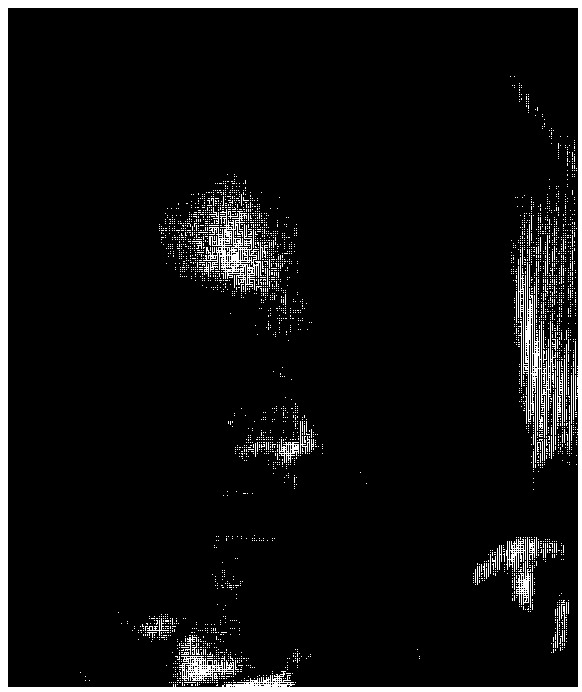
[0002] At present, the three-dimensional imaging of fetal body surface structure can only be obtained by three-dimensional or four-dimensional ultrasound imaging. figure 1 It is the fetal facial image obtained by three-dimensional ultrasound; the technical method is single, and the ultrasound imaging conditions are affected by many factors such as fetal position, structural occlusion, and amniotic fluid volume, and sometimes it is impossible to obtain a satisfactory three-dimensional image of the fetal body surface
In addition, the positive findings of ultrasonography also lack the confirmation and support of other imaging examinations
[0003] Magnetic resonance scanning is safe, radiation-free, and has higher tissue resolution. It has been widely used in the diagnosis of fetal prenatal diseases. However, due to the characteristics of its imaging signals, it is difficult to mask the surface structure of the fetal body during its three-dimensional post-processing. Imaging and maximum intensity projection methods, so it is difficult to obtain three-dimensional images of fetal body surface structure based on magnetic resonance examination by conventional methods

Method used


Image

  • Visual three-dimensional imaging method for fetal body surface structure based on magnetic resonance scanning
  • Visual three-dimensional imaging method for fetal body surface structure based on magnetic resonance scanning
  • Visual three-dimensional imaging method for fetal body surface structure based on magnetic resonance scanning

Examples


Embodiment Construction

[0019] The following will clearly and completely describe the technical solutions in the embodiments of the present invention with reference to the accompanying drawings in the embodiments of the present invention. Obviously, the described embodiments are only some, not all, embodiments of the present invention.

[0020] refer to Figure 2-5 A method for visualizing three-dimensional imaging of fetal body surface structure based on magnetic resonance scanning proposed by the present invention, comprising the following steps:

[0021] S1 Fetal magnetic resonance data acquisition, fetal magnetic resonance data acquisition can be carried out with various types of existing magnetic resonance scanners, and technicians can master the technical essentials of scanning after simple training.

[0022] MRI scanning and data acquisition requirements:

[0023] MRI scanning sequence: real steady-state free precession gradient echo sequence, which is equipped with all models of mainstream m...


PUM


Abstract

The invention discloses a visual three-dimensional imaging method for a fetal body surface structure based on magnetic resonance scanning. The method comprises the following steps of S1, collection offetal magnetic resonance data; S2, data conversion; S3, three-dimensional post-processing; and S4, three-dimensional imaging for the fetal body surface structure, completing volume rendering displayby use of a ray-casting method so as to calculate a color value at a pixel point on a screen. According to the visual three-dimensional imaging method for the fetal body surface structure based on magnetic resonance scanning, the three-dimensional data collection for a fetal-interested region organization structure is performed by use of a real steady-state free precession sequence and an existingmagnetic resonance imaging hardware facility, new data containing fetal histological characteristics are generated through data processing, surface reconstruction is performed on the new data by useof three-dimensional post-processing software to obtain a three-dimensional image of the fetal body surface structure.

Description

technical field [0001] The invention relates to the technical field of medical imaging, in particular to a three-dimensional imaging method for visualization of fetal body surface structure based on magnetic resonance scanning. Background technique [0002] At present, the three-dimensional imaging of fetal body surface structure can only be obtained by three-dimensional or four-dimensional ultrasound imaging. figure 1 It is the fetal facial image obtained by the current 3D ultrasound; the technical method is single, and the ultrasound imaging conditions are affected by many factors such as fetal position, structural occlusion, and amniotic fluid volume, and sometimes it is impossible to obtain a satisfactory 3D image of the fetal body surface. In addition, the positive findings of ultrasonography also lack the confirmation and support of other imaging examinations. [0003] Magnetic resonance scanning is safe, radiation-free, and has higher tissue resolution. It has been w...

Claims


Application Information

Patent Timeline
Patent Type & Authority Applications(China)
IPC IPC(8): G06T17/00G06T15/08A61B5/055A61B5/00
CPCA61B5/055A61B5/72G06T15/08G06T17/00G06T2200/08
Inventor 肖连祥张翼丁蕾魏新红林祥涛
Owner 肖连祥
Learn More