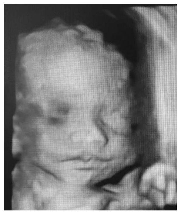
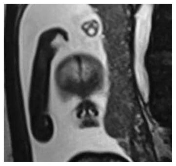
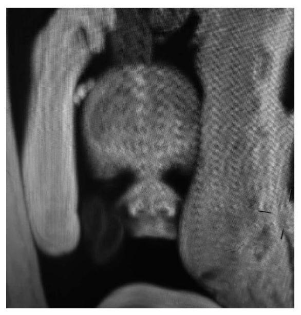
Three-dimensional imaging method for visualization of fetal body surface structure based on magnetic resonance scanning
A magnetic resonance scanning and three-dimensional imaging technology, which is applied in the field of medical imaging, can solve the problems of not being able to obtain a satisfactory three-dimensional image of the fetal body surface, lack of confirmation and support from other imaging examinations, and obtaining the structure of the fetal body surface, so as to achieve excellent image quality, The effect of intuitive image and broad application prospects
- Summary
- Abstract
- Description
- Claims
- Application Information
AI Technical Summary
Problems solved by technology
Method used
Image

Examples
Embodiment Construction
[0019] The following will clearly and completely describe the technical solutions in the embodiments of the present invention with reference to the accompanying drawings in the embodiments of the present invention. Obviously, the described embodiments are only some, not all, embodiments of the present invention.
[0020] refer to Figure 2-5 A method for visualizing three-dimensional imaging of fetal body surface structure based on magnetic resonance scanning proposed by the present invention, comprising the following steps:
[0021] S1 Fetal magnetic resonance data acquisition, fetal magnetic resonance data acquisition can be carried out with various types of existing magnetic resonance scanners, and technicians can master the technical essentials of scanning after simple training.
[0022] MRI scanning and data acquisition requirements:
[0023] MRI scanning sequence: real steady-state free precession gradient echo sequence, which is equipped with all models of mainstream m...
PUM
 Abstract
Description
Claims
Application Information
 - R&D
- Intellectual Property
- Life Sciences
- Materials
- Tech Scout
- Unparalleled Data Quality
- Higher Quality Content
- 60% Fewer Hallucinations
Browse by: Latest US Patents, China's latest patents, Technical Efficacy Thesaurus, Application Domain, Technology Topic, Popular Technical Reports.
© 2025 PatSnap. All rights reserved.Legal|Privacy policy|Modern Slavery Act Transparency Statement|Sitemap|About US| Contact US: help@patsnap.com