Blood optical-detection method and device

An optical detection and blood technology, applied in the direction of Raman scattering, material excitation analysis, etc., can solve the problems of pain in blood component detection, long waiting time for test results, complicated test methods, etc., to achieve improved comfort, strong pressure, and detection method fast effect

Inactive Publication Date: 2018-05-18
武汉比色光电科技有限公司
View PDF7 Cites 1 Cited by
  • Summary
  • Abstract
  • Description
  • Claims
  • Application Information

AI Technical Summary

Problems solved by technology

Existing testing methods for blood components are painful, there is a risk of infection, and the waiting time for test results is long
Moreover, the existing detection methods have different problems and bottlenecks such as high false positive rate, complicated test methods, expensive instruments, and long time-consuming.

Method used


Image

  • Blood optical-detection method and device
  • Blood optical-detection method and device
  • Blood optical-detection method and device

Examples


Embodiment 1

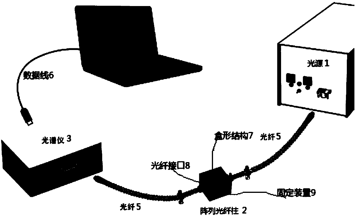
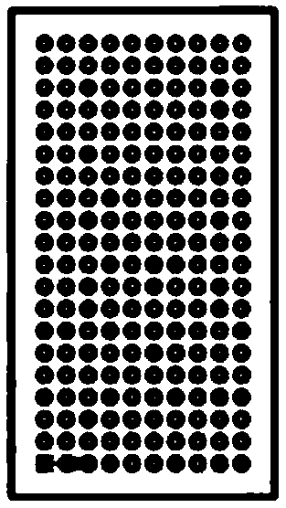
[0036] Such as figure 1 As shown, a blood optical detection device includes a light source 1 , a microarray fiber column 2 , a spectrometer 3 , an optical fiber 5 , a data line 6 and a computer 4 . Both the micro-array fiber column 2 and the light source 1 , and the micro-array fiber column 2 and the spectrometer 3 are connected through the optical fiber 5 . The spectrometer 3 is connected to the computer 4 through the data line 6 . The micro-array fiber column 2 is installed on the transverse card slot through rows and rows of installation holes. A box-shaped structure 7 with an opening at the bottom of the box is arranged on the periphery of the micro-array fiber column 2 , and the box-shaped structure 7 is used to cover the micro-array fiber column 2 . Both sides of the box-shaped structure 7 are provided with optical fiber interfaces 8 for connecting the optical fibers 5 . The box-shaped structure 7 is provided with a fixing device 9 for fixing on the human body during ...


PUM


Abstract

The invention discloses a blood optical-detection device and method. The blood optical-detection device comprises a light source, a miniature array optical-fiber column, a spectrometer, optical fibers, a data line and a data processing end, wherein the miniature array optical-fiber column consists of paired miniature optical-fiber needle columns; each miniature optical-fiber needle column is provided with a reflecting film and a light-transmitting film; the optical fibers are respectively adopted for connection between the miniature array optical-fiber column and the light source and between the miniature array optical-fiber column and the spectrometer, and the spectrometer is connected with the data processing end by the data line. The method using the device to detect blood comprises thefollowing steps that the light source emits light, the light enters the blood by one miniature optical-fiber needle column to generate a spectral Raman effect with components of the blood, and then outgoes by another miniature optical-fiber needle column, the spectrometer splits the outgoing light, and then converts light signals after light splitting into electric signals, and the data processing end carries out analysis and calculation on the electric signals by a mathematical model and obtains a detection result.

Description

technical field [0001] The invention relates to a blood optical detection method and device. Background technique [0002] The content of blood components can reflect certain physical conditions of the human body. Conventional blood component detection and analysis mostly requires the collection of blood samples by pricking the skin of the finger tip or drawing a large amount of blood from a vein, and then obtaining the values ​​of biochemical indicators in the blood through spectroscopic or chemical analysis. Blood component detection and analysis methods mainly use microscopes to check the types and sizes of cells in blood, gas chromatography liquid chromatography and mass spectrometry and other spectroscopic optical techniques, and enzyme-linked immunosorbent assays. Existing testing methods for blood components are painful, there is a risk of infection, and the waiting time for test results is long. Moreover, the existing detection methods have various problems and bot...

Claims


Application Information

Patent Timeline
Patent Type & Authority Applications(China)
IPC IPC(8): G01N21/65
CPCG01N21/65
Inventor 张峰胡良锦张垒
Owner 武汉比色光电科技有限公司
Why Eureka
Try Eureka