Eureka AIR delivers breakthrough ideas for toughest innovation challenges, trusted by R&D personnel around the world.

Brain tumour image segmentation method and device based on improved full convolutional neural network

A convolutional neural network and image segmentation technology, applied in the field of medical devices, can solve problems such as low efficiency and instability, and achieve the effects of improved accuracy, strong practicability, and low computer memory overhead

Inactive Publication Date: 2018-03-02
TIANJIN UNIV
View PDF3 Cites 41 Cited by
  • Summary
  • Abstract
  • Description
  • Claims
  • Application Information

AI Technical Summary

Problems solved by technology

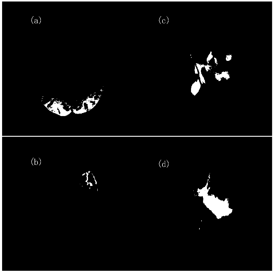
[0006] In order to overcome the deficiencies of the prior art, the present invention aims to propose an improved fully convolutional neural network to realize the machine segmentation of brain tumor MRI images, avoid the defects of low efficiency and instability in manual segmentation, and provide fast and reliable The segmentation results of brain tumors can provide accurate basis for the diagnosis, treatment and surgical guidance of brain tumors.

Method used


Image

  • Brain tumour image segmentation method and device based on improved full convolutional neural network
  • Brain tumour image segmentation method and device based on improved full convolutional neural network
  • Brain tumour image segmentation method and device based on improved full convolutional neural network

Examples


Embodiment Construction

[0039] The invention combines medical images and deep learning algorithms to complete the segmentation of brain tumor nuclear magnetic resonance images. This fully automatic brain tumor image segmentation will have an important impact in the field of medical imaging.

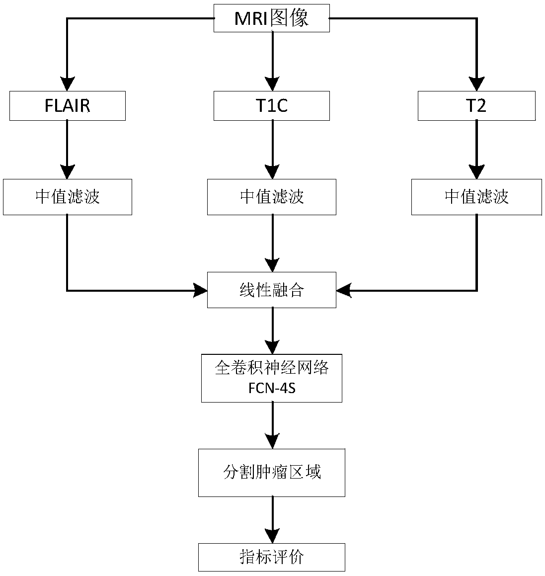
[0040] Aiming at the defects of the traditional convolutional neural network in image segmentation, the present invention proposes an improved fully convolutional neural network, which is successfully applied in the segmentation of brain tumor MRI images, avoiding the low efficiency of manual segmentation and instability defects. Using a new deep learning algorithm to provide fast and reliable brain tumor segmentation results, thus providing an accurate basis for the diagnosis, treatment and surgical guidance of brain tumors.

[0041] In order to achieve the above object, the present invention adopts the following technical solutions:

[0042] 1) Select an image. The quality of the MRI brain tumor image itsel...


PUM


Abstract

The invention relates to the medical apparatus field, aims to propose an improved full convolutional neural network to realize machine segmentation of a brain tumor nuclear magnetic resonance image, avoids low efficient and instability existing in manual segmentation, provides the rapid and reliable brain tumor segmentation result and provides accurate bases for diagnosis, treatment and operationof brain tumors. The method comprises steps that 1), an image is selected; 2), a full convolutional neural network FCN model is constructed; 3), the segmentation result is tested, when the FCN model is trained, the trained model is utilized to predict the tumor position and the boundary size of any brain tumor image, corresponding evaluation indexes are utilized to evaluate the segmentation result, and the FCN model can be better ameliorated. The method is advantaged in that the method is mainly applied to NMR image processing occasions.

Description

technical field [0001] The present invention relates to medical devices, in particular to a method and device for segmenting brain tumor pictures based on an improved full convolutional neural network. Background technique [0002] The brain is the most important part of the human body, but the incidence of brain tumors has been on the rise in recent years. According to surveys, the number of brain tumors diagnosed in the United States increased by 23,000 in 2015 alone. The World Health Organization divides brain tumors into five grades according to the degree of lesions. Brain tumors are mainly divided into two types: benign tumors and malignant tumors. Benign tumors such as meningioma can generally recover after surgical treatment, while gliomas and gelatinous tumors can generally recover. Malignant tumors such as mesenchymal tumors are difficult to cure because of their stubbornness, so they are also called brain cancer. Magnetic resonance imaging (Magnetic Resonance Ima...

Claims


Application Information

Patent Timeline
IPC IPC(8): G06T7/11G06T5/50G06T3/40
CPCG06T7/11G06T3/4007G06T5/50G06T2207/10088G06T2207/20032G06T2207/20081G06T2207/20221G06T2207/30016G06T2207/30096
Inventor 邢波涛李锵关欣
Owner TIANJIN UNIV
Why Eureka
Eureka Blog
Learn More